Pages

Wednesday, 30 November 2011

What's in the box? No.7 - loadsa leaf-beetles and an interloper

As promised, I'm back to my pet theme - beetles - in particular, the mysterious specimens that arrive by post because I coordinate the UK's Chrysomelid Recording Scheme which covers what are commonly known as leaf, reed, seed, tortoise & flea beetles (i.e. the Chrysomelidae, Bruchinae, Donaciinae, Zeugophorinae, Megalopodidae, Cassidinae and Orsodacnidae). So, now you are sufficiently overloaded with taxonomic terms (not to mention the fact that whether or not some are considered families or subfamilies varies by book/website), let's have a look at what prompted me to start writing...


What arrived in the post...
Usually I get a single specimen tube with one or two beetles in, but not this time... What I have here is an old Kodak slide-box full of about 20 little envelopes, each of which has several beetles of different species in. The envelopes are neatly labelled (date, location, UK grid reference) and the beetles look to be in pretty good condition. Also, the sender did get in touch first (and included return postage so he can have the specimens back once identified) so I expected more than the usual number of specimens. Most will be 'flea beetles' (subfamilies Galerucinae & Alticinae) as they are the ones the sender couldn't identify himself, and these are the ones most likely to be problematic (even to specialists on occasion). They also represent the parts of my test key which are receiving the most comprehensive rewrite as some of the keys to these genera didn't work well enough. So, although it will take a long time (it looks like there are about 100-200 beetles), it's really useful to have some unknown specimens to work through while I rewrite the relevant keys, as well as providing useful data for the recording scheme. It will also give me plenty to blog about... so, on to the first envelope!

A closer look at the contents of the first envelope.
Here it is clear that two of the beetles are larger (around 8mm long) and less smooth and shiny. These are reed beetles (subfamily Donaciinae) and will be the subject of a separate post soon. The one that stands out - to me anyway - is the small black roundish one more-or-less in the middle of the group.

A close-up of that round blackish beetle.
This beetle is around 3mm long and although shiny, you can see tiny punctures on the surface of the wing-cases (elytra). However, this isn't what grabbed my attention - it's the leg sticking out, in particular the pair of spurs at the joint of the femur and tibia. This gives me a good idea of what this beetle is, but let's turn it over to make sure.


Ventral view showing the enlarged hind femurs
Close-up of the enlarged hind femur. Personally I like the detail of small hairs in this photo.
The large femurs enable this beetle to jump and similar structures are seen in the 'flea beetles' within the family Chrysomelidae. However, flea beetles don't tend to be this round, nor do they have leg-spurs quite like those seen here; also, although you can't see the detail here, some fine details of the structure of the head are different from the chrysomelids. In fact, this isn't a chrysomelid at all - it is Scirtes hemisphaericus which is in the family Scirtidae and, as noted by Cooter & Barclay (2006), is sometimes confused with the flea beetles because of its legs and jumping ability. It's also in a family that is relatively unfamiliar, possibly because there is no modern book covering the British Scirtidae; however, the key in Joy (1932) still works well (although it was then called the Helodidae) and is available on a CD-ROM which is rather more affordable than the original 2-volume book (as it happens, I have the 1976 reprint which wasn't too expensive as collectors don't like ex-library books because of the stamps and marks - personally I don't care as long as it's complete).

I intend to continue this series, especially as I should have no shortage of material, interspersed with other ecological musings. In this case, I've started the 'mystery chrysomelid' thread with something that isn't a chrysomelid at all - I wonder what will appear next...

References

Cooter, J. & Barclay, M.V.L. (eds.) (2006). A Coleopterist's Handbook (4th ed.). Amateur Entomologists' Society, Orpington. An absolute must for beetle enthusiasts! Based on the UK beetle fauna.
Joy, N.H. (1932). A Practical Handbook of British Beetles (2 vols.) (1976 reprint). Classey, Farringdon.

Tuesday, 22 November 2011

What's in the box? No.6 - taxonomic confusion and big shoulders.

OK, after my brief foray into palaeontology, it's time to get back to what I arguably know a bit more about - tiny invertebrates. You may have seen previous posts like this one and this one about the specimens that land on my doormat having been sent to me for identification as the organiser of the UK Chrysomelid Recording Scheme. I usually know that something is on its way as I ask for a little warning from contributors to make sure I don't get sack-loads of beetles when I'm busy, on holiday etc, especially as the organiser role is voluntary... This case was no exception as the collector had been in touch to ask about a specimen that appeared to be Aphthona ?atratula. Now, the ? in the name isn't a typo - it's there to indicate that there is some taxonomic confusion surrounding this species in Britain. Basically, A. atratula was referred to as A. atrovirens Foerster, 1849 by Pope (1977) but this species is not found in Britain (Cox, 2000). This means that British specimens of 'A. atrovirens' are probably A. atratula Allard, 1859. So, that's actually quite straightforward isn't it... well, actually, no. The problem is that various authors in continental Europe (e.g. Konstantinov, 1998) consider A. atratula to be the same as A. euphorbiae (Schrank, 1781), the Large Flax Flea Beetle. However, there are some quite convincing differences between British specimens of A. atratula and A. euphorbiae suggesting that they are actually separate - including the spread of A. euphorbiae with cultivated flax which is not seen in A. atratula (Cox, 2007). I happen to agree that they are separate species but officially this is not certain and so the ? remains. In any case, that's enough of the finer points of Chrysomelid taxonomy for now; let's have a look at the specimen itself.

Dorsal view of the Aphthona specimen, length 2mm excluding appendages. This is how it arrived in the post - very neatly (unlike my own card-mounting), glued onto a small card in a protective tube - thanks to Jon Cole for the good quality specimen!
First of all, it is an Aphthona - although I didn't get a clear photo of it, there is a spur on the outer side of the lower edge of the hind tibia (honest) - if it was in the middle of the lower edge, it would be in the genus Phyllotreta - see why the 'flea beetles' are considered so tricky to identify! I'm not going to go through the whole process of keying out Aphthona specimens here, but let's look at some of the final features required to make a species-level identification.

A close-up of the front half in dorsal view.
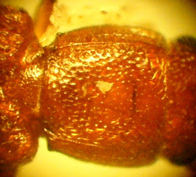
Looking at the pronotum, you can see some small 'punctures' - these are actually moderately coarse as finer ones would look more-or-less like a matt surface. So, it can't be A. melancholica which has very fine punctures - it is, as expected, either A. atratula or A. euphorbiae (remembering that Konstantinov, 1998 and several other important authors would stop at this point, considering the species to be the same). Both can be shiny black with a bluish reflection as seen here, however do have a look at the 'shoulders' of the elytra (wing cases). Here, they are well developed and are somewhat bulbous - this is characteristic of A. euphorbiae as is the size with A. atratula reaching no more than 1.7mm and having weakly developed shoulders. This is a fairly good indication that the specimen is actually of the common and widespread A. euphorbiae, but isn't strictly conclusive - to be really certain, dissection of the male genitalia is required (as is so often the case with small chrysomelids), and handily, this is a male.

Ventral view of the aedeagus.

Dorsal view of the aedeagus.

Side view of the aedeagus.
These images can be compared with those here at the excellent 'European Chrysomelidae' website. They clearly belong to A. euphorbiae - the dorsal view for example bears the same small polygonal structure at the base of the indent at the tip and the side view shows the same curvature. A. atrovirens (remember the taxonomic confusion...) however does not show the same small polygonal structure and the curvature differs slightly in that the inside edge of the curve straightens towards the tip.

So, we have a common species, but one that provides an opportunity to work through the taxonomy of Aphthona. Though often associated with cultivated flax/linseed (Linum usitatissimum) which they may kill by feeding on seedlings and cotyledons (Cox, 2007), it is actually found on many plant species in a wide range of habitats - as in this case where the beetle was collected (by beating in late October) from a Field Maple (Acer campestre) by a lake. Now, a box of about 20 little packets of beetles has arrived so I suspect this series will be updated soon...

References

Cox, M.L. (2000). Progress report on the Bruchidae/Chrysomelidae Recording Scheme. The Coleopterist 9: 65-74.
Cox, M.L. (2007). Atlas of the Seed and Leaf Beetles of Britain and Ireland. Pisces, Newbury.
Konstantinov, A.S. (1998). Revision of the Palaearctic Species of Aphthona Chevrolat and Cladistic Classification of the Aphthonini (Coleoptera: Chrysomelidae: Alticinae). Associated Publishers, Gainesville, FL.

Monday, 21 November 2011

The eyes have it: Trilobites as models of ecology and evolution

Following my recent scribblings about cave bear dentition, I thought I would make the journey back to looking at extant invertebrates a step at a time. So, staying in the world of palaeonotology, but moving onto invertebrates (slightly more familiar ground), I decided to see if I could derive some more inspiration from my curio shelves. So, given that I've previously written about my Cretaceous water bug, it seemed about time I tackled that most popular of fossil invertebrates, the trilobite. Now, I've only got the one trilobite, but it's quite a good specimen, so here it is in all its glory:

My trilobite, 5cm long and showing clear segmentation plus its well preserved head on the right.
The shape of this, with a bulging and pimply glabella ('forehead') suggests it is in the genus Phacops (possibly P. rana) but not Reedops as this would have a smooth glabella. However, what brings me straight to the suborder Phacopina in the first place is its eyes (see Murray 1985 for a key to trilobite groups).

Phacops compound eye showing clearly separated lenses. This is the right eye looking from above and slightly off to the right. The pimply surface to the left is the fixed 'cheek' area known as the fixigena which appears as a lobe on either side of the glabella.
Trilobite eyes are compound like those of modern invertebrates (well, up to a point) but vary greatly, and this one is known technically as 'schizochroal'. Only found in the Phacopina, and not all of these, eyes of this type have relatively few relatively large lenses as can be seen in the photo. These lenses are well separated and if we took a section through the eye, apart from destroying my trilobite, we would see that each lens had its own cornea (outer layer - which we also have) and was separated from the others by a thick piece of exoskeleton called the sclera with the cornea extending down through this. This is not the case in most trilobites which have 'holochroal' eyes. These have small lenses which are often more numerous and may look more like our familiar 'mosaic' picture of an insect's eye. The lenses all touch each other (i.e. are not separated by sclera) and have a common cornea covering the whole surface of the eye. Lastly, a few trilobites (only the Cambrian Eodiscina) have 'abathochroal' eyes which have few, small lenses which are separated as in schizochroal forms but only have thin sclera and a cornea which stops at the surface of the sclera. Got all that? Well, I've only just worked through the anatony of these eye types and found an excellent summary on S.M. Gon's webpage 'The Trilobite Eye' which I recommend if you would like a expanded and illustrated version of my account here as well as other variant such as stalked eyes and those with inbuilt eyeshades!

Apart from my liking for all things morphological, one important aspect of these eye forms (and those trilobites that were eyeless) is what they can tell us about the life/ecology of trilobites. For example, eye loss is seen in some benthic (bottom-feeding) forms which lived in low-light conditions. So, starting with Phacops, it has fully developed eyes and can be seen as an ancestral form of genera with reduced eye development such as Cryphops which in turn evolved into the eyeless Trimerocephalus. So, we have a genus with well developed eyes which evolved into forms reducing and then losing them - a process that took a long time in human terms but occurred in the, er, blink of an eye, when looking at geological timescales (see Dawkins 1996 for more on the evolution of eyes of various types).

This loss of eyes in a benthic environment is a simple enough concept, but what about the development of schizochroal eyes in the first place? Unlike most (holochroal) trilobite eyes they are highly specialised and have no clear analogue in the modern fauna (Fortey, 2000). Firstly the lenses are crystalline, being made of calcite and are almost spherical, sometimes a little drop-shaped. These lenses have even had photographs taken through them and it is evident that sharp images could be formed and that larger 'pieces' of the trilobite's surroundings would have been visible per lens than for those with holochroal eyes. However, trying to use spherical transparent items such as marbles in a visual system doesn't work well because of 'spherical aberration' - the images become distorted, inverted, fuzzy. However, Phacops solved this problem by making the calcite impure, specifically by replacing some of the calcium atoms in calcite with magnesium and forming an internal 'bowl' in the lens which worked as a corrective structure, separating the eye into two sections of differing refractive index and allowing for the spherical aberration (Clarkson & Levi-Setti, 1975). So, although holochroal eyes would presumably have been good at detecting movement (food, predators?) as is the case in many modern invertebrates, Phacops could see chunks of detail. It is unknown exactly why this type of eye evolved, but it arose, like all other evolved structures, because it improved survival, in this case through a process known as post-displacement paedomorphosis (i.e. the retention of juvenile features - part-developed holochroal eyes are like smaller versions of schizochroal ones).

So, there we have it - Phacops evolved a visual system which has not (yet) been 'repeated', but why did I call trilobites 'models of ecology and evolution' in the title of this article? Well, the evolutionary side is well documented (despite what hordes of frankly bizarre creatonist and Intelligent Design websites might assert to the contrary - however, I won't go there...) both in the scientific literature and in popular-science publishing/broadcasting - despite the hundreds of millions of years that separate trilobites from our modern Earth, they are to some extent familiar. As for the ecology, it allows us to mix some evidence with a dash of speculation. Benthic lifestyles with low light levels led to the loss of eyes and so we can infer something of the 'lifestyles' of genera such as Trimerocephalus from their morphology as well as from the location and material in which they are found. However, in Phacops, we have some evidence of what level of detail they might have been able to see - I say might because their optic nerves are not preserved, thus their 'wiring' remains a mystery as far as I am aware, and by extension so is precisely how they perceived the world.

I'll stop there - I hope you enjoyed that, and please do watch this space for a return to the wonderful world of small beetles soon!

As in some modern invertebrates, the rear segments formed a section behind the thorax known as the 'pygidium' AKA 'the end'!

References

Clarkson, E. N. K. & Levi-Setti, R. (1975). Trilobite eyes and the optics of Des Cartes and Huygens. Nature 254: 663-667.
Dawkins, R. (1996). Climbing Mount Improbable. Viking, New York.
Fortey, R. (2000). Trilobite! Eyewitness to Evolution. Flamingo, London.
Gon, S.M. (2007). The Trilobite Eye. http://www.trilobites.info/eyes.htm [accessed 20/11/2011].
Murray, J.W. (ed.) (1985). Atlas of Invertebrate Macrofossils. Longman, London.

Tuesday, 15 November 2011

The Truth of the Tooth: Cave Bear dentition, ecology and extinction

Lately I have written copiously about small invertebrates, particularly those found recently in our firewood store. So, having written five parts of the woodpile series (so far), I felt it was time for a brief departure - in terms of both time and scale as I have decided to look at some aspects of the Cave Bear Ursus spelaeus.

A Cave Bear skeleton in the typical (of museum displays) rearing posture.
Cave Bears lived in Pleistocene Europe (the Pleistocene epoch lasted from around 2.5 million to 11,700 years ago and covers the most recent series of repeated glaciations) and current evidence suggests they became extinct around 27,800 years ago. This means that they would have been encountered by humans and indeed they are depicted in cave art (albeit rarely e.g. at Les Combarelles cave in France). There is also possible evidence of Cave Bear worship by Neanderthals, such as at Drachenloch in Switzerland and Regourdou in France where the skulls of bears had clearly been arranged in and on man-made stone structures such as wall niches and a slab-covered pit. However, prehistoric anthropology, fascinating though it is, really isn't my area, so I'll stick with the more biological/ecological aspects. However, for an interesting overview of some aspects of human-Cave Bear interactions (focusing more on the earlier form of Cave Bear U. deningeri which disappeared around 100,000 years ago and may be an earlier species, a transitional subspecies or simply a pre-interglacial form of U. spelaeus), have a look at Stiner (1999). Taxonomic uncertainties aside, my interest was sparked when I bought a Cave Bear cheek tooth found in a cave in Romania, and dental evidence is where I will start.


Cave Bear cheek tooth, length 45mm.
This tooth is in pretty good condition (it's still shiny after about 40,000 years which shows just how tough tooth enamel is) and has an extensive grinding surface with a couple of large bluntly pointed cusps. Cave Bears lost their premolars as they evolved, a feature which has been used to suggest a highly herbivorous diet (e.g. Kurtén, 1976). The last premolar evolved as a molar (molarisation) which allowed tough plant material to be chewed more effectively (and hence more more food energy to be extracted) due to the increased number of cusps and cutting edges of the teeth, especially in the elongated last molar. Their teeth also show more wear than in most modern bears which again suggests a herbivorous diet with a large component of tough/fibrous materials, although detailed analysis indicates that tubers and other gritty foods were not a major part of their diet, unlike for modern Brown Bears U. arctos (Pinto Llona et al., 2005). However, varying threads of research in this area, including evidence for some cannibalistic scavenging (Pacher, 2000) has led to current scientific opinion tending towards Cave Bears being more herbivorous than modern bears of the genus Ursus, but still omnivorous to some extent. Recent re-examination of skull and tooth morphology (Figueirido et al., 2009) and analysis of the regional variation in bone isotope composition, especially nitrogen-15 (Richards et al., 2008; Trinkaus & Richards, 2008) both support this idea of omnivory and some variation in diet.

The same cheek tooth showing the pattern of the crown. The large grinding surface covers the left side of the tooth and the lower right side, with the pointed cusps to the upper right. The orange deposits in the grooves of the enamel are the remains of soil, although the right-hand end shows an area of worn (yellowish and not shiny) enamel at the base of the large cusp.
With advances in molecular biological techniques, the possibility of investigating cave bear genetics arose and in 2005, nuclear DNA extracted from a Cave Bear tooth around 42-44,000 years old was sequenced. This indicated that the Cave Bear was more closely related to the Brown Bear and Polar Bear U. maritimus than to the North American Black Bear U. americanus (Noonan et al., 2005) and supported earlier similar findings using mitochondrial DNA (Loreille et al., 2001). Interestingly, investigation of the fine structure of Cave Bear tooth enamel (the 'rods' or 'prisms' that form the basic units of enamel) shows that it retained carnivore-like characteristics despite the clear adaptation to a largely herbivorous diet. Thus, changes in broad dental anatomy driven by dietary specialisation can occur without the equivalent changes in enamel structure (von Koenigswald, 1992), meaning that Cave Bears had herbivore-shaped cheek teeth with carnivore-like enamel.
So, we have an extinct species of bear clearly adapted to a specialised herbivorous diet with some elements of omnivory and variation. As well as the genetic evidence mentioned above, its skeleton is similar to that of the modern Brown Bear, with the two species appearing to have diverged around 1.2 to 1.4 million years ago (Loreille et al., 2001) i.e. prior to the splitting of Brown and Polar Bears which may have occurred around 850,000 years ago, although this estimate is somewhat uncertain (Swenson, 2007). Males averaged 400–500 kg with females around half this weight at 225–250 kg (Christiansen, 1999), similar to the range for the largest modern bears, noting that they were larger during glaciations and smaller during interglacial periods (MacDonald, 1993), probably as an adaptation to adjust heat loss rate as larger animals have smaller surface area:volume ratios. The reason for its extinction is uncertain. It is unlikely to simply be due to its specialised diet and restricted geographical range ecologically 'marooning' it during post-glocial warming - after all, it had survived several similar changes in condition previously and there is possible genetic evidence of a decline starting some 25,000 years prior to its extinction (Stiller et al., 2010). Also, as noted above there is strong evidence for the species' ability to vary its diet. Instead, it is likely that there was a complex interplay of factors, possibly involving competition with humans for cave habitat, maybe specifically for hibernation sites as Cave Bears did not appear to use alternatives such as forest thickets and failure to find a hibernation site would lead to death. Despite numerous media reports taking the 2010 paper by Stiller et al. to be definitive evidence of competition with humans rather than changing climatic conditions to be the cause of Cave Bear extinction, there is still genuine scientific disagreement and research is ongoing. Further genetic work (Bon et al., 2011) does however show reduced genetic diversity from specimens in France originating from the period directly prior to extinction (genetic diversity is greater for specimens prior to this), again indicating a species under stress during human colonisation of the area - and the possibility of competition for hibernation caves.


References

Bon, C., Berthonaud, V., Fosse, P., Gély, B., Maksud, F., Vitalis, R., Philippe, M., van der Plicht, J. & Elalouf, J.-M. (2011). Low regional diversity of late cave bears mitochondrial DNA at the time of Chauvet Aurignacian paintings. Journal of Archaeological Science 38 (8): 1886-1895. 
Christiansen, P. (1999). What size were Arctodus simus and Ursus spelaeus (Carnivora: Ursidae)? Annales Zoologici Fennici 36: 93–102.
Figueirido, B., Palmqvist, P. & Pérez-Claros, J.A. (2009). Ecomorphological correlates of craniodental variation in bears and paleobiological implications for extinct taxa: an approach based on geometric morphometrics. Journal of Zoology 277 (1): 70–80.

Kurtén, B. (1976). The Cave Bear Story. Life and Death of a Vanished Animal. Columbia University Press, New York.
Loreille, O., Orlando, L., Patou-Mathis, M., Philippe, M., Taberlet, P. & Hänni, C. (2001). Ancient DNA analysis reveals divergence of the cave bear, Ursus spelaeus, and brown bear, Ursus arctos, lineages. Current Biology 11 (3): 200203.
MacDonald, D. (1993). The Velvet Claw: A Natural History of the Carnivores. BBC, London.

Noonan, J.P., Hofreiter, M., Smith, D., Priest, J.R., Rohland, N., Rabeder, G., Krause, J., Detter, J.C., Pääbo, S. & Rubin, E.M. (2005). Genomic Sequencing of Pleistocene Cave Bears. Science 309 (5734): 597599.
Pacher, M. (2000). Taphonomische Untersuchungen der Höhlenbären-Fundstellen in der Schwabenreith-Höhle bei Lunz am See (Niederösterreich). Beiträge zur Paläontologie 25: 11–85.
Pinto Llona, A.C., Andrews, P. & Etxeberrıa, P. (2005). Taphonomy and Palaeoecology of Cave Bears from the Quaternary of Cantabrian Spain. Fondacion de Asturias/Du Pont Iberica/The Natural History Museum, Grafinsa, Oviedo.
Richards, M.P, Pacher, M., Stiller, M., Quilès, J., Hofreiter, M., Constantin, S., Zilhão, J. & Trinkaus, E. (2008). Isotopic evidence for omnivory among European cave bears: Late Pleistocene Ursus spelaeus from the Peştera cu Oase, Romania. Proceedings of the National Academy of Sciences of the United States of America 105 (2): 600604.
Stiller, M., Baryshnikov, G., Bocherens, H., Grandal d'Anglade, A., Hilpert, B., Munzel, S.C., Pinhasi, R., Rabeder, G., Rosendahl, W., Trinkaus, E., Hofreiter, M. & Knapp, M. (2010). Withering Away 25,000 Years of Genetic Decline Preceded Cave Bear Extinction. Molecular Biology and Evolution 27 (5): 975978.


Stiner, M.C. (1999). Cave bear ecology and interactions with Pleistocene humans. Ursus 11: 4158.
Swenson, J.E. (2007). Økologi hos en voksende bjørnebestand – Forvaltning når bjørnen har kommet tilbake. Det Skandinaviske Bjørneprosjektet [in Swedish] [accessed 15/11/2011].

Trinkaus, E. & Richards, M. P. (2008). Reply to Grandal and Fernández: Hibernation can also cause high δ15N values in cave bears. Proceedings of the National Academy of Sciences of the United States of America 105 (11): E15.
von Koenigswald, W. (1992). Tooth enamel of the cave bear (Ursus spelaeus) and the relationship between diet and enamel structures. Annales Zoologici Fennici 28: 217227.

Monday, 14 November 2011

There's a Black Widow in my shed (not)!

Recently, while writing one of my series about woodpile invertebrates, an Australian reader/blogger mentioned the rather larger and more dangerous invertebrates found down under - here in the UK, most invertebrates are harmless to humans and even those that can bite or sting rarely cause major problems unless you are allergic to them. So, when I found a particular spider in my shed, I was reminded of occasional media reports of people finding, or even being bitten by, 'black widows'. Now, we don't have black widows (genus Latrodectus) here, but we do have 'false black widows' (Steatoda) which are also in the family Theridiidae and look superficially similar. Spiders in this family are known collectively as the comb-footed spiders due to the bristles on the tips of their hind legs which they use to tease out silk to form 'tangle' - rather than sticky - webs.

My Steatoda specimen (about 9mm long) showing the bulbous abdomen typical of the Theridiidae - also note the short hairs. The pattern shows a pale, ywllowish arc and spots on a purplish background.
The features above show this to be S. grossa - a female - although the pattern can be highly variable or even absent; Roberts (1993) provides detailed diagrams of female epigynes and male palps if required for identification. This species is found mainly in and around houses in southern England (sometimes in coastal areas of the south-west) and although traditionally considered quite scarce, it appears to be increasing in range and frequency due to the warming effects of climate change. A similar increase has been seen in the non-native S. nobilis (NHM, 2007).

The Natural History Museum (NHM) receive enquiries about bites from Steatoda species, but spider-bites are uncommon in the UK (just a few reported each year); only 12 species are capable of biting humans (including the two Steatoda mentioned so far) out of a total of about 640 species. No-one has ever died of a spider-bite in the UK, and serious effects are rare (to be honest, true black widow bites are only occasionally fatal, though they are very painful and unpleasant). So, what should you do if you find a spider like Steatoda in the UK? Well, apart from temporarily incarcerating this specimen in order to take photos (it's now back in the shed along with other specimens, inlcuding males), I tend to leave them alone. If you want to handle them, it is easy to be careful by collecting them in a suitable container e.g. if you want to put them outdoors (or into the shed). Personally, I'm happy to leave them be, especially as I generally only see them when moving things around in the shed, at which point they flee and hide. Happy spidering!

Here's looking at you - a dorsal view of the pearly eyes of S. grossa.


References

Natural History Museum (2007). The truth about false widow spiders.[accessed 14/11/11]. Lots of snippets of info about this group of spiders in the UK.
Roberts, M.J. (1993). The Spiders of Great Britain and Ireland (compact ed.) (2 vols.). Harley, Colchester. This is the standard comprehensive work on spiders of the British Isles and is excellent, but it isn't cheap! There is a 2009 reprint by Apollo Books.

Wednesday, 9 November 2011

Bark at the Moon - small invertebrates of timber (Part 5)

More from the smaller end of the macroinvertebrate scale... I got distracted from writing a book about some larger beetles (Chrysomelidae) and went back to the woodpile critters. Yesterday, I posted some images of the beetle Cryptolestes duplicatus and mentioned in passing that it was a parasite/predator of other beetle larvae. I didn't really elaborate on this at the time, so I decided to take another shot to get an image of the head and go from there.

The head of C. duplicatus showing the mandibles


As you can see, the mandibles are well developed as are the eyes so, despite being associated with bark (within a genus often associated with stored food products and thus seen as pests), it clearly isn't always in dark conditions - it may not be as fearsome as, say, a tiger beetle (Cicindelidae), but it appears well equipped to hunt. Although there is a lot of literature on Cryptolestes covering the pest status of constituent species and various taxonomic revisions, there doesn't appear to be much on its biology and ecology. However, Lukin (2010) does note that the larvae of C. duplicatus are fungus feeders beneath bark during the early stages of the decomposition of coarse dead wood.

Moving onto a species I haven't mentioned yet in this series, one other beetle caught my attention today, in particulaer the neat arrangement of hars on its dorsal surface...

Another small beetle - note the neat rows of long hairs and the curved ridges running parallel to the sides of the pronotum.
Similar in size to the previous species (around 1.7mm long), this was a relatively easy identification as I was familiar with its photograph in Hurka (2005), in particular, the pronotal ridges, long hairs and oval form. It is a specimen of Mycetaea subterranea and is in the family Endomychidae, close relatives of the ladybirds (Coccinellidae) - they are sometimes called the 'false ladybirds'. This particular species is sometimes known as the Hairy Cellar Beetle or the Handsome Fungus Beetle and is most often associated with cellars and outbuildings (barns, stables etc), although it is sometimes found in rotten wood inside hollow trees. Both larvae and adults feed on fungi.

That is enough for today - as mentioned before, I intend to keep working on the woodpile invertebrates and have some as-yet unidentified mites, barklice and, yes, beetles to work on, so this series isn't finished yet!

References

Hurka, K. (2005). Beetles of the Czech and Slovak Republics. Kabourek, Zlin.
Lukin, V. (2010). Species structure of the saproxylic beetles assemblages in the protected territories of Belarus.Muzeul Olteniei Craiova. Oltenia. Studii şi comunicări. Ştiinţele Naturii 26(2): 155-160.

Tuesday, 8 November 2011

Bark at the Moon - small invertebrates of timber (Part 4)

After yesterday's brief journey off-topic (sometimes it just has to be done), I'm back to the tiny invertebrates found in our firewood store. As promised, I've started looking at the sub-2mm critters I must say are proving tricky. This is not because of their small size as such - after all, I have microscopes - but because they include taxa which are relatively unfamiliar, not only to me, but given the lack of literature covering some of them, to entomologists more broadly. There are also difficulties associated with handling them without damage (tiiiiny tweezers, small brushes) and storing them without some of the soft-bodied organisms shrivelling to almost nothing. This has as much to do with my lack of curatorial expertise with such species as anything else. However, there is an up-side; these issues probably mean there are some under-recorded species among the bark and dead-wood (saproxylic) invertebrate community and hence some interesting records if I can make species-level determinations. First up, another barklouse or 'psocid'.


A head-on view of a barklouse. The tiny size is highlighted by using the individual lenses (ommatidia) of the eyes as a rough idea of scale.
Although it can't be seen in the photo above (due to the specimen having shrivelled I think - it's still there but very faint), there is a diagnostic anchor-shaped mark on the 'face'. The wings are reduced to tiny buds and the abdomen has several rows of dots, still visible and appearing as round 'bumps' here, though their bumpiness is a bit of an optical illusion. This is the widespread Cerobasis guestfalica which has been spread internationally through commerce such as the timber trade. Almost all specimens found are females (males occur very very rarely) which means most poulations are entirely parthenogenetic i.e. they reproduce without fertilisation by a male (New, 2005). Moving back into my comfort zone - beetles - I managed to find a single specimen of a 1.6mm bark beetle.


Note the elongate shape, rounded antennal segments and longitudinal lines on the elytra and pronotum.

A close-up of the pronotum. The hind (left) angles have a tiny tooth and there are two slightly raised lines on each side of the pronotum - the inner one is quite clear, the outer one less so, though it can be seen as a broken bright line.
This beetle is in the family Laemophloeidae - not a group I am very familiar with - and to identify it, I needed to go back to Joy (1976). The features above did however allow identification as Cryptolestes duplicatus, a species which is probably predatory and/or parasitic on the larvae of other beetles. This species has a scattered, localised distribution in south and south-east England; not rare as such but not common either, and I suspect under-recorded (as well as having seen some significant taxonomic changes which have moved it from the genus Laemophloeus within the family Cucujidae), so quite a good find. Feeling bold, I thought I'd move onto something really small - a mite (so, an arachnid rather than an insect).

A mite showing the hard surface with a few long hairs (and some bits of plant material stuck to it!), but no velvety covering as seen in the more familiar 'spider mites'.
For now, I'm not going to try to identify this - I don't have much literature covering the mites, though I do think it is in the family Acaridae, based on its overall form. However, if I feel keen I might try later, in which case an update will appear here.

So, what next for the 'Bark at the Moon' series? Well, I still have some specimens to identify and I intend to continue collecting, so although posts in this series may slow down a bit, I strongly suspect there are more to come. After all, how else to investigate these under-recorded groups..?

References

Joy, N.H. (1976). A Practical Handbook of British Beetles (2 vols.). Classey, Faringdon. (This is the resized reprint of the original 1932 classic work). A CD-ROM is also available.
New, T.R. (2005). Psocids. Psocoptera (Booklice and Barklice). RES Handbooks for the Identification of British Insects 1(7): 1-146.

Monday, 7 November 2011

Why physics says convenience culture is bad for the environment

I imagine some regular readers will have seen the title of this post and thought, "what?", "that's not what the Ecology Spot is about - what's Dave up to?" Well, I agree it is a bit of a departure from the norm - I generally keep away from the big science-and-philosophy topics because there are plenty of bloggers out there already doing just this, and in many cases doing it very well. However, an idea popped into my head and sometimes such ideas just have to be followed to see where they go... So, where did it come from?

Well, when I'm not scrutinising invertebrates and other organisms, I sometimes like to leave my comfort zone and delve into a bit of maths and physics - usually somewhere in between the paperbacks-for-beginners (too basic) and textbooks (too hard). To be honest, this is a fairly narrow line for a writer to tread, so there aren't that many books to choose from, but there are some - Feynman, Greene, Hawking, Penrose... ah, Penrose... You see, I've been reading his new book, Cycles of Time and very interesting it is too. Most of it isn't relevant here, but I got to one section (pages 77-79 in the 2011 paperback) which explained that the energy the Earth receives from the sun more-or-less equals the energy it radiates. I knew this (amaong other things, I teach courses covering climate change, so ought to really), and I knew that the 'yellow' photons from the sun have a higher frequency that the infra-red ones the Earth emits. What I had not realised was that this means that the sun provides the Earth with energy in a lower-entropy form than that which it emits. Que? Well, the overall amount of energy emitted equals that received but as this energy is spread across a greater number of lower-frequency photons, the entropy ('randomness') increases upon emission - there are more photons, hence more 'degrees of freedom'.

Now, you might quite rightly be wondering where all this is going and what it has to do with convenience culture. The answer is 'nothing' directly, but it is what started me thinking (always a risk) about topics like conservation of energy and so on. Without going into exactly how I got there, the key thought that appeared from all this was essentially:

"If 'convenience' means doing things without putting any personal effort in, where does the energy to do them come from?"

See what I mean? If I want to dig a hole in my garden, some resources are used to manufacture my shovel and provide me with food as fuel for my labours. However, if I decide that I want the hole without the labour, any other option (apart from not creating the hole) uses more resources. If I get a machine, this will have been manufactured using more resources than the shovel, and will need fuel (I will need to eat either way). An operator or delivery person may need to be paid and this money comes from my income which in turn, somewhere along the line, derives from the use of natural resources from a finite system. So, the more I spend (i.e. consume), the more resources are being depleted somewhere (unless all resources are fully renewable, which they aren't). Given how our economy works (or is meant to - it seems to be wobbling rather a lot at the moment), this ultimately derives from an environmental good of some sort - timber, oil or whatever - the impacts of which (deforestation, climate change, other pollution) tend to be treated as 'externalities' in economics i.e. they are not included as intrinsic costs. If they were, cheap disposable items would be a lot more expensive. The same goes for 'convenience' food and anything else which replaces our physical effort with work done by a device or process.

Now, I'm sure I'm not the first to notice this and I already know that extra packaging and energy use are involved in consumer goods (let alone the uselessness of many such goods in the first place) and more so if they are disposable, but it is the first time I've really thought about the fundamental physical inevitability of 'convenience' - we get nothing for free and if we don't do the work ourselves (whatever it may be), finite resources elsewhere have to do it for us, with the resulting impacts that entails. Of course, the above is greatly simplified - there are variations according to where my food comes from, machine efficiency and so on, and those involved in calculating carbon footprints, life-cycle analyses and so on will be familiar with such details. However, precise figures aside, 'convenience' = extra resource use.

OK, enough from me - I hope I stopped before descending into soapboxing waffle. Now those thoughts are out in the open, I shall be returning to the usual ecology next post...

Reference

Penrose, R. (2010). Cycles of Time. Vintage, London.

Friday, 4 November 2011

Bark at the Moon - small invertebrates of timber (Part 3)

OK, only an hour to go before I head off to the firework display at our local pub, so apologies for any typos that get missed in the hurry...

Anyhow, as promised I've started looking at some of the tinier invertebrates found in our firewood store - by tinier I mean below 2mm but still visible (just) with the naked eye. I have quite a few still to look at, but the first is a springtail (Collembola) which despite its small size can be identified without a microscope if you have good close-up vision.


A springtail - the head is to the right and the 'spring' is out of frame to the left.
This springtail is Entomobrya albocincta and can be identified by the 'mane' of straggly hairs which also run along the dorsal surface, and the alternating dark and pale patches which give it a striped appearance. Even without a microscope, the palest mark just behind the head is clearly visible and the other mark just behind the mid-point can also be seen. This is a common and widespread species (there were plenty in our wood store) and is indeed usually associated with dead wood or found under bark (Hopkin, 2007).

As I mentioned above, I've only just started looking at the smaller residents of our wood store and I'm sure there are other sub-2mm specimens I'll be able to identify. However, along the way I found a couple of other interesting species, a little larger at 3-4mm.

An unusual creature - very flat with short wing-buds. Also note the pattern of red spots near the rear end. The overall colour and pattern provide excellent camouflage on bark and among lichen and plant material.

The 2nd and 3rd antennal segments are the same length. Note also the spines either side of the rostrum and the red eyes.

A close-up of the midline of the dorsal surface showing tiny pinkish tubercles (bumps) and other sculpturing.
This slightly odd-looking specimen is a nymph of one of the flatbugs, Aradus depressus, a true bug (Hemiptera) in the familt Aradidae. Adults develop fully sized wings and this species is often found on the damp ends of cut timber - no great surprise to find it among firewood. It is again a fairly common species and feeds on the mycelia (threads) and fruiting bodies of Polyporus and other fungi (Southwood & Leston, 1959).Moving on, I found what looked like another barklouse (psocid)...

A barklouse with short, hairy/scaly wings and long antennae. The bulging eyes suggest it is a male.

A close-up of the head - note the clear pattern of dark marks. The dense layer of silky hair-like flattened scales can be seen covering the wings and there are also larger bristles around the outer edge. The antennae have numerous small segments, each with its own small hairs.
This combination of features is quite distinctive and shows this to be a specimen of Pteroxanium kelloggi. It is usually found in leaf litter and is only rarely seen on bark or attached foliage (New, 2005), so is not usually associated with timber piles. It is the only species of the family Lepidopsocidae regularly found outdoors in Britain - there are three other species of this family known from Britain, but they are rare, casual introductions. The name of the family is also a good indicator of its form in this case - like the Lepidoptera (scaly-wings) i.e. the butterflies and moths, it is named after the dense flat scales on the forewings.

And so, we have some more species from our wood pile - it is turning out to be a local biodiversity hot-spot! It's time for the weekend to start now, but I should return soon with Part 4 of this series and hopefully more on the tinier residents.


References

Hopkin, S.P. (2007). A Key to the Collembola (Springtails) of Britain and Ireland. FSC, Shrewsbury.

New, T.R. (2005). Psocids. Psocoptera (Booklice and Barklice). RES Handbooks for the Identification of British Insects 1(7): 1-146.
Southwood, T.R.E. & Leston, D. (1959). Land & Water Bugs of the British Isles. Warne, London.

Tuesday, 1 November 2011

Bark at the Moon - small invertebrates of timber (Part 2)

After yesterday's look at a species of barklouse (Psocoptera) which hasn't been known from Britain for very long, I thought I'd move on to the invertebrate group arguably most often associated with dead wood - beetles.I have so far only worked on the 'larger' (2-3mm) specimens that I collected late last week from our firewood store; tinier species remain to be identified...So, I shall begin with a broadly familiar group, the weevils (Curculionidae).

Side view showing punctures, cylindrical shape and broad, blunt rostrum.

Head showing rostrum (without a strongly broadened end or sharp basal excision) and bluntly pointed (rather than rounded) ends to the antennae.

Rear of elytra (wing cases) showing a flange round the edge (technically, formed by  outgrowth of the 9th interstice or 'gap between rows of punctures').
Using the excellent work my Morris (2002) for this often fiddly group, these features lead to identification as Euophryum confine, a now-common species in Britain; introduced here from New Zealand in 1937, it has spread rapidly and can be a pest of timber (including buildings). I'll be checking our firewood carefully before bringing it indoors!

Next, a beetle with a broadly similar size and overall form (unsurprising for wood-borers that have to fit into tunnels and small holes) - despite only being 2-3mm long, this one was quite strikingly red in colour even to the naked eye.

Dorsal view showing reddish colour and cylindrical shape.

Ventral view of the head and thorax. The spines at the front corners of the pronotum are clearly visible, as are its smooth (rather than toothed) sides. Also, the 'temples' of the head are not sharply toothed.

Dorsal view showing that the pronotum does not have a pair of longitudinal grooves.
Again, this combination of features shows it to be a specimen of Silvanus unidentatus with Hurka (2005) proving useful for separating it from the rarer S. bidentatus. This is another cosmopolitan species which can be a timber pest. However, where there are small invertebrates, there are of course others for whom they are a food source. Some insectivores are well-known - many birds feed on small insects, and some of course break into wood to find them. However, among the small invertebrates of timber, there are similarly sized predators, in this case one of the true bugs (Hemiptera).

A bug with one of the fairly typical hemipteran body shapes, more or less droplet shaped with a pointed head widening evenly to the abdomen. This specimen is short-winged ('brachypterous') and the legs are yellowish with darkened femora.

Looking more closely, the wings are clearly brownish. Note the typical hemipteran scutellum - the triangular area between the wings and the pronotum.
This time using an older publication, Southwood & Leston (1959), it was fairly straightforward to key this specimen as Xylocoris cursitans, a bug in the family Cimicidae, the group which includes the bed-bugs (which have a similar general form to this species). A widespread species in Britain, it lives under the bark of fallen trees and is predatory on small beetles, springtails and thrips, and so may well help to control potential timber pests such as those described above. Of course, being a bug-nerd, I find the 'pest' species interesting in their own right, but I still have no intention of inviting them in to eat our tasty floorboards!

So, with the larger (relatively speaking) specimens covered, the next stage of my timber investigations will be to look at some of the tinier inhabitants to see what else lurks among the bark and wood-fibres. Part 3 hopefully on its way soon...

Part of our firewood store - it has since expanded considerably - what else will be calling it 'home'?

References

Hurka, K. (2005). Beetles of the Czech and Slovak Republics. Kabourek, Zlin. An excellent and well-illustrated overview of beetles from this part of Europe; the majority of species covered are found in Britain.
Morris, M.G. (2002). True Weevils (Part I). Coleoptera: Curculionidae (Subfamilies Raymondionyminae to Smicronychinae). Handbooks for the Identification of British Insects 5(17b): 1-149. Currently the standard work for this group in Britain.
Southwood, T.R.E. & Leston, D. (1959). Land & Water Bugs of the British Isles. Warne, London. Though old, this is still very useful and is available as a 2005 reprint (and possibly CD-ROM) from Pisces Publications, which is the version I have - the original is not cheap!