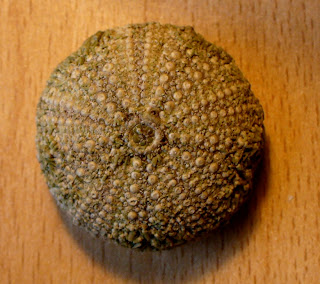
So, starting at the start, the ID was straightforward enough. The specimens were all 25-40mm in diameter, somewhat flattened on top and greenish with spines (of those still bearing spines) fairly short, mostly of about the same length, and green with purplish tips. This, along with investigation of the plates and pores, makes them specimens of Psammechinus miliaris, a common species around the coasts of Britain (Hayward & Ryland, 2000).
 |
| 40mm specimen with spines |
 |
| 25mm specimen without spines |
 | |
| Basal joint/attachment of spines. |
 |
| Greek columns? No, a garden of spines |
Looking at the top, the periproct (anal area) and surrounding plates can clearly be seen. There is a large plate (the 'madreporite') with numerous small pores and one larger gonopore, plus four other genital plates, each also with a gonopore - as the names suggest these are involved in reproduction.
 |
| The periproct, surrounded by four genital plates and the larger madreporite (upper right). |
 |
| Above and below, mysterious pink ovals |
Though tiny, they look almost like little shells, but for now, they are known as UPOs (unidentified pink objects). So, dissecting pin in hand, I managed to extract one (it was quite well attached) and put it under the microscope. This is what I saw...
 |
| Top image x40, middle pair x100, bottom image x400 |
First of all, I must admit I had no real idea what the UPO might be. It looks like a tiny bivalve or maybe brachiopod, but I've no clue about the size or form of, say, early post-larval bivalves, even of common species. It appears to have an attachment structure and threads at what I'm currently calling the 'hinge' or 'base'. A join/suture separates two 'valves'; this is straight basally, and sinuous apically. There is coarse surface sculpturing, although this is fine on the paired bulbous structures.
*** UPDATE - MYSTERY SOLVED ***
I posted images on iSpot and sent some to the Marine Biological Association (MBA), in the hope of more information appearing soon - and iSpot did its job after just a day. I had wondered if it was something unusual, or something very familar to marine invertebrate specialists, but otherwise not so widely known about. It was even suggested that it might be a seed blown onto the stranded urchin as there are plant seeds with sculpturing not unlike this. However, an iSpot user came up with a much more straightforward suggestion which turns out to be correct - it's the tip (or 'business end') of a pedicellaria, and so part of the urchin after all. I had been completely thrown by the unfamiliar structure without a stalk, but of course in hindsight it's obvious - the stalks seen all over the surface have in many cases got flat ends because the tips have become detached. There are few good images around showing this structure, but once I knew what I was looking for, I came across this site (the only one so far) with excellent photos, including the whole structure - the text is in Japanese, but the images do their job.
So, after all this detective work, I remain fascinated - I generally work on terrestrial and freshwater species and had no idea that these structures existed (it might have been mentioned in Zoology 101 way back, but that was a while ago...) - small mobile jaws and poison darts: wonderful! It still makes sense for a small organism to hide among urchin spines - fossil brachiopods have even been found on fossil urchins in the US (Schneider 2003). This is the sort of location where it is quite possible no-one ever looks, so even something common may well be under-recorded and such places (like the holdfasts of seaweeds) remain a potential source of interesting finds. It's also a reminder to assume nothing!
References
Hayward, P.J. & Ryland, J.S. (eds.) (2000). Handbook of the Marine Fauna of North-West Europe. OUP, Oxford. [a great book if you can get it].
Schneider, C.L. (2003). Hitchhiking on Pennsylvanian Echinoids: Epibionts on Archaeocidaris. Palaios 18(4-5): 435-444.


