Welcome

Welcome to my blog

This is where I post various musings about wildlife and ecology, observations of interesting species (often invertebrates)
and bits of research that grab my attention. As well as blogging, I undertake professional ecological & wildlife surveys
covering invertebrates, plants, birds, reptiles, amphibians and some mammals, plus habitat assessment and management
advice
. I don't work on planning applications/for developers. The pages on the right will tell you more about my work,
main interests and key projects, and you can follow my academic work here.
Showing posts with label galls. Show all posts

Tuesday, 4 February 2014

Mystery gall time

January was quiet on the blogging front - a combination of grotty weather and mucho other work. However, I'm back with a mystery gall sent to me by Phil Budd from the Southampton Natural History Society (although he found it in Enfield, Greater London). He found it (with about 50 others) in a sallow tree, Salix cinerea and had noted the presence of what looked like moth larvae living in/on it. Beyond this, identification remained elusive and so it was sent to me for further scrutiny.

Gall (approx 20mm diameter) on a Salix cinerea twig - it has clearly developed on one side of the twig and has a swollen and irregular surface.
One side of the gall was softer with fibrous material, possibly the remnants of a galled bud.

My first stop was the excellent Redfern & Shirley (2011), a standard (and affordable) work with excellent coverage of British galls, though the rapidity of change in cecidology (gall study) means here are always new species being added and new host-galler association being found. It quickly became clear that this is either a species not found in that book, or an unfamiliar form of a gall which is included. Either way, it was time to open the gall and look at the the larvae.

Larva (approx 12-14 mm long) from the Salix cinerea gall.
Opening the gall showed that there were several larvae inside and that they were still alive. I removed one which, from the arrangement of three pairs of true legs towards the front, a gap in the middle, then prolegs towards the rear, was indeed a moth larva. It has a darkened head capsule and first segment, bristles and a pinkish-brown colour, with paired dots on the segments in dorsal view.

Larva in dorsal view, noting the paired dots clearest on the front few segments.
This looks (to me) very much like the larva of Cydia servillana, a moth in the family Tortricidae which, although uncommon, is known to cause galls on this tree species. However, the gall is an elongate spindle-shaped swelling nothing like the knobbly and irregular gall seen here. It also contains a single larva, unlike the case here where there were several larvae together. So, what are the possibilities?

  • It could be C. servillana creating an unusual form of gall, or utilising an existing growth of the type sometimes caused by the tree's response to a wound of infection.
  • It could be a different species of moth which I can't find reference to or which hasn't been recorded before, at least not as a galler of S. cinerea.
  • An unknown gall causer such as that noted on catkins in Redfern & Shirley (2011).I have my money on this option...
  • Something I haven't thought of. Also distinctly possible!
Although I couldn't identify this for certain, there were some things I could do...

  • As the larvae are alive, try to raise them as adults and identify the moths that emerge.
  • Ask a gall-specialist - in this case I forwarded this post to the British Plant Gall Society.
As it happens the BPGS responded very quickly and confirmed one of my maybes/suspicions - it is probably the unknown call-causer, maybe a virus or phytoplasma (specialised bacteria that parasitise the phloem and are transmitted by sap-feeding insects much as malaria is transmitted by mosquitos) that distorts catkins, and the larvae are simply using the structure for shelter. So, the gall itself remains a mystery, but the moth may be identifiable if it develops to adulthood - if so, I'll post an update. Until then, you can see that the larva I removed is alive and well...

The gall is held above the larva which then climbs onto it and begins to investigate the various holes and crevices.
The larva continues to explore.
After a few minutes, the larva entered the gall - hopefully it will pupate and emerge as an adult.

Reference

Redfern, M. & Shirley, P. (2011). British Plant Galls (2nd ed.). FSC, Shrewsbury.

Sunday, 22 September 2013

Dining on smut and other corny puns

A couple of weeks ago, I wrote about the smut fungus Ustilago maydis that grows on sweetcorn. While doing that, I found out that it is edible, and in Mexico is eaten as a delicacy called 'huitlacoche'. I also mentioned that if I found some more that wasn't quite so over-mature, I'd try it. I found some, I cooked it, and I ate it. So, in a rare departure from ecology on 'the spot', here are a couple of pics of the taste test.

A cob with U. maydis growing on it, being chopped and prepared.
Bits of huitlacoche fried and ready to eat.
The outcome of the taste test was pretty favourable - unsurprisingly a kind of cross between mushroom and sweetcorn, and quite subtle. I've found another growth in our sweetcorn patch, so that will be cultivated and I'll try more next time...

Sunday, 8 September 2013

Smutty farming

Yup, an obvious pun I know, but hey... as you may know if you read this blog regularly, I am a stakeholder in a community farm - we're chemical-free and pretty wildlife-friendly, and as such use either (a) cunning or (b) many pairs of hands to deal with pests. Of course, we get a few, but they can be interesting in their own right - like yesterday when I found a single fist-sized growth of common smut fungus (Ustilago maydis) at the base of one of our sweetcorn plants.

Common smut Ustilago maydis on sweetcorn
Close-up of U. maydis showing a small growth of the sweetcorn plant.
The fungus can grow on any part of the plant, though, as here, it most commonly affects the cob/seeds which expand and become filled with spores - essentially it causes a gall. It is fairly common, and its presence this year is unsurprising as conditions have been ideal - hot, dry weather while the plants are establishing, followed by rain as they mature - exactly what has occurred. It can cause major crop losses, though our non-intensive 'hands-on' approach means that we'll simply look out for and remove any more if they appear (the standard advice is to burn or bin them, but not compost them as the spores will survive and spread).

Interestingly though, I have found out that the fungus is actually edible, particularly if relatively young (this specimen was very mature and entirely spore-filled), and in Mexico is a delicacy known as 'huitlacoche' which is eaten in a succotash, or in tacos or omelettes. Apparently it's kind of nutty-mushroomy and quite nice but not popular elsewhere as it's seen as a 'disease'/'rot' rather than food - and I must admit it doesn't look that appealing. However, if I find another, less mature one, I'm going to try it - seems a good use of a failed cob! I may report the results here...

Friday, 27 April 2012

Who you gonna gall?

If you are interested in plant galls, you'll know how important it is to correctly identify the host plant as there is often a high degree of specificity between the gall host and the gall causer which makes identification a lot easier. It's not always that straightforward however, as I discovered when I found galls on the leaves of a Mountain (or Alpine) Currant Ribes alpinum.

Galls on a leaf of Ribes alpinum
These are clearly true galls as the red patches are swollen rather than simply being discoloured, and looking Redfern & Shirley (2011) - the standard work on British galls - there are few options on Ribes. In fact, the key quickly moves to an answer, both caused by aphids of the genus Cryptomyzus:

  • On redcurrant R. rubrum, galls caused by C. ribis which are yellow-green in colour.
  • On R. alpinum, galls caused by C. korschelti which are pink, orange or reddish.
The plant ID is definitely correct (a known specimen confirmed by an experienced botanist), so it looks like C. korschelti, but it's important to check carefully, so let's see the aphids themselves.

Aphids on the underside of an R. alpinum leaf

Close-up of an aphid on the underside of an R. alpinum leaf
By now you should have seen the difficulty - this is a yellow-green aphid, but this implies the aphids are C. ribis which are not, according to Redfern & Shirley (2011), found on R. alpinum. However, rather than having found a new species, I thought it was more likely that this is R. ribis on an unusual (for the UK) host. However, having consulted with an aphid specialist (thanks Fiona!), not only is it unusual to find Cryptomyzus on the underside of the leaf rather than inside the galls, but the galls themselves are too swollen. So, let's look even more closely...

Siphons (or 'cornicles') at the rear of the aphid's abdomen
Cornicles are an important way to separate some aphid species, and these are broadly swollen with a slightly widened rim at the end (this isn't very clear in the photo, but it is there), the whole being approximately bottle-shaped. In Cryptomyzus, the cornicles are narrower - this is a different genus. Without going into too much detail here, it turns out that (thanks again Fiona) it is in the genus Hyperomyzus, specifically H. lactucae. This is rarely recorded in Britain (though this doesn't necessarily mean it is rare, just that not many people look or can identify them), but it has been found here before and is known from Ribes in continental Europe.

Many galls are not well understood and minor discoveries like this can be made quite readily if care is taken to look, especially given that R. alpinum is not especially common in the UK and probably poorly studied. The lack of readily accessible/affordable identification guides (Blackman & Eastop's 2006 2-volume opus was needed for this species, but is not cheap) makes aphid study more difficult (plus some genera are taxonomically confusing and really require genetic analysis and/or research), but as I found, there are specialists who are ready to help and it's always worth asking. Now to let the authors of the gall key know what I've found, then check whether H. lactucae has been recorded in Hampshire before...

References

Blackman, R.L. & Eastop, V.F. (2006). Aphids on the World's Herbaceous Plants and Shrubs. (2 vols.). Wiley, Chichester.
Redfern, M. & Shirley, P. (2011). British Plant Galls (2nd ed.). FSC, Shrewsbury.

Thursday, 15 March 2012

Oh, my rusty hollyhocks!

As my wife knows, I don't have to be at work to get distracted by an interesting bug, plant or fungus. So, nobody was very surprised when, during a leisurely walk following a family pub lunch yesterday, I bent down to take a sample of an interesting-looking hollyhock (Alcea rosea) leaf.

Hollyhock leaf showing orange fungal structures and something small indicated by a red arrow...
As you can see from this photo, the leaf bears numerous orange fungal structures on both the blade and petiole (stalk), plus there's a small invertebrate indicated by the red arrow. Staring with the fungus, a closer look highlights some familiar structures...

Elongate orange fungal structures on the petiole. These are blister-like and the dark orange masses are spores that have become exposed as the blisters have ruptured.
On the leaf blade, similar (but more globular) spore-filled structures are seen (red arrows), while some appear greyer in colour (blue arrow).
Although small, these blisters clearly include some abnormal growth and colouration of the plant's tissues and thus can be considered to be galls. A quick look in Redfern & Shirley (2011) provides an easy identification - there is a single galling species species found on hollyhock (and some other Malvacaeae), the rust fungus  Puccinia malvacearum. This specimen clearly matches the description, including the grey colour of older spores. Although this species may be unfamiliar, the genus is found on many plants with those on lords-and-ladies (Arum maculatum) and nettles (Urtica dioica) being particularly common and widespread - just look out for the little orange rings and pustules. Now onto the potentially trickier tiny invertebrate...

Small (3 mm long) leafhopper on the hollyhock leaf. See below for the meaning of the red arrows and circle.
If you are familiar with generalist insect books such as Chinery (1986), you can quickly tell that this is a leafhopper (Cicadellidae) of some sort, possibly a relative of the often-illustrated Eupteryx aurata. However, there are numerous species in this group (the subfamily Typhlocybinae) and the more technical Le Quesne & Payne (1981) may well be needed to identify them by keying out. So, back to a bit of taxonomic morphology - important diagnostic features are as follows:

  • The three apical forewing veins (indicated by red arrows in the above photo) join the same cell (indicated by the red circle), noting that two of the veins merge to form a 'Y'.
  • In the same photo, you can see the white 'waxy area' on the front edge of the forewing. Just behind thisthere is an irregularly shaped black spot cut into two by a pale wing vein. In some species this spot marges into one.
  • In side view the front of the face (running down to the piercing mouthparts) is flat without a clear angle part of the way down (see top photo below).
  • The pattern on the pronotum is distinctive - two clear black dots near the front edge with fuzzy longitudinal brown marks attached to them, plus other smaller black dots to the sides (see lower photo).
  • The pattern on the head shows a triangle of three large black spots; the single rear spot does not have a dent in its front edge.
  • The front of the 'face' is not clearly shown here, but in the above photo you can just see that there are two more black spots in front of the three on the head, but not another pair further to the side by the eyes.
Side view of the leafhopper showing the flat front to the 'face'.
More-or-less dorsal view of the leafhopper showing the patterns on the head and pronotum.
Combining these features and using them in the key, the species can be identified as Eupteryx melissae. This is a leafhopper which, like the rust fungus above, is known from hollyhocks and related Malvaceae. It is very similar to E. thoulessi but can be separated using the features on the front of the face (E. thoulessi has the extra pair of lateral spots near the eyes which are absent here), and of course the food-plant is a helpful clue. Although this specimen was seen in mid-March, it is usually not recorded until May. However, it may have emerged following a recent spell of warm weather; when collected it appeared dead, but was probably simply torpid as the temperature had fallen considerably over a couple of days - certainly it became active again under the microscope.

More soon...

References

Chinery, M. (1986). Collins Guide to the Insects of Britain and Western Europe. Collins, London.
Le Quesne, W.J. & Payne, K.R. (1981). Cicadellidae (Typhlocybinae) with a checklist of the British Auchenorhyncha (Hemiptera, Homoptera). Royal Entomological Society Handbooks for the Identification of British Insects 2(2c): 1-95.
Redfern, M. & Shirley, P. (2011). British Plant Galls (2nd ed.). FSC, Shrewsbury.

Thursday, 27 October 2011

Rollin' rollin rollin' - the diverse inhabitants of a willow leaf

Over the last year or so (yup,  the 'Ecology Spot' is nearing its first birthday!) I've posting several musings on the inhabitants of galls - not only the gall causers but also other species that can make galls their home. However, there is something conceptually similar, and closely related biologically, that I haven't looked at yet, and that is leaf-rollers.

Leaf-rollers, as the term suggests, are organisms that roll leaves to form shelters - unlike galls, the plant doesn't grow new structures, although there is some overlap (excuse the pun) with some leaf-rolls including further distortions such as thickening and increases in cell number, and hence being considered galls (Redfern & Shirley, 2011). In this case, the roll in a willow (Salix) leaf (found at Winnall Moors on the edge of Winchester, southern England) involves twisting and thickening of the plant tissues and so is classed as a gall (only just - sometimes the thickening is very slight).

Willow leaf roll showing twisting and thickening (increased cell size is visible in places)
 The downwards roll affects both sides of the leaf and most of the blade which indicates that it was caused by one of the Phyllocolpa species, a genus of sawflies in the family Tenthredinidae. Indeed, there were small dark sawflies with yellowish legs in the area which may well have been Phyllocolpa, although I did not manage to catch a specimen (either physically or photographically). As the larvae of these sawflies drop to the ground to pupate in the soil, it is not uncommon for galls to be empty, but the only way to find out is to look...

The roll unrolled - at first glance there is nothing but frass (invertebrate faeces)

Looking more closely, a few pale creamy-white rounded-oval eggs can be seen, each no more than 0.5mm in diameter. Whatever they are, an adult invertebrate was here recently. We'll return to these later...

A small cocoon of tangled silk. The lid (top right) is detached showing that whatever developed here has now left.

The bottom of the cocoon showing attachment threads plus a dark patch that may be frass.
So, we have eggs and an empty cocoon - is there anything more immediately identifiable? Well, fortunately yes. Whether or not they are in any way related to the cocoon I don't know, but two exuviae ('skins') were also present.
An unidentified exuvium - probably one of the Hemiptera (true bugs) with clearly defined wing buds and abdominal segments.

That's more like it - something recognisable, an exuvium of a small spider; even the leg hairs are clearly visible.
Although the 'bug' had long gone, the spider (or at least a spider) remained.

Dorsal view of the tiny spider (a few mm long) found in the leaf roll.

A close-up showing various appendages and some of the eyes.
Although the abdominal pattern seems quite clear, this is probably a juvenile. From the general form, I suspect it is one of the orb-weavers (Araneidae) though I would be more than happy if an arachnologist could clarify! If it is this family, it may be using the leaf roll for shelter as hunting takes place on an orb-web (a small one may have been present but un-noticed of course). Along with the spider I also found the following insect already dead (the abdomen was dry and wrinkled) - I wonder if it had become spider-food?

Ventral view of the insect showing the pointed 'snout', long antennae and spotted wings held in a tent-like position.

Dorsal view of the head showing protuberant compound eyes and orange simple eyes (ocelli). Note the patterning on the head and the bristly antennae.
This is a psocid or barklouse - the tented wings with spots at the tips of the wing veins, plus the habitat (on  foliage of trees and shrubs) make this a straightforward identification as the genus Ectopsocus. It is probably the common E. briggsi, but dissection is needed to separate it from E. petersi and E. meridionalis with ceryinaty, and the taxonomy of these closely related species remains uncertain (New, 2005). The protuberant, almost stalked, eyes mean it is probably a male.

And so, that brings us to the end of the specimens that could be identified (to some extent at least) at the time of collection about two weeks ago. However, I did mention that we would return to the eggs. Once found, I put them on their section of leaf in a small container to see if they would hatch, and they did...

Tiny larva about 1mm long - dorsal view

Ventral view showing segmented legs.

Ventral close-up of the head and thorax.
The dorsal view looks superficially somewhat like a woodlouse (i.e. it is 'onisciform'), but the darkened and hardened ('sclerotised') head with small pointed mandibles, plus the legs, show that it is actually a tiny beetle larva, one of two that hatched. It may not be immediately obvious, but this is actually a larva of a 'flea beetle', one of the small species of jumping beetles, the tribe Alticini within the family Chrysomelidae, although it might be within the wider subfamily Galerucinae within which the Alticini are included. The legs are short and have 4 segments (with a claw) and the mandibles are simple and sickle-shaped without a grinding surface ('mola'). There are thin bristles around the body (e.g. just visible top left in the 3rd photo) but no long cerci (tail-like appendages), the antennae are tiny and conical, and willows are a favourite host plant (Cooter & Barclay, 2006). Having just hatched, this is a first-stage larva and hence difficult to identify further, though van Emden (1942) is a standard work that is often useful for larval identification to family. However, the Macro-invertebrate Lab at the City Valley State University have kindly produced a Digital Key to the Aquatic Insects of North Dakota. They have good clear images of a more fully developed larva of this type, and also images of larva at genus level for Pyrrhalta - very similar to what was found here.

So, at present, I have identified all the organisms within this single leaf roll as far as I can. I hope it shows the importance of such small-scale habitats and the diversity they support, as well as highlighting some groups that are likely to be under-recorded due to their small size and tendency to hide. These under-recorded groups and habitats are worth taking the time to investigate as there are discoveries to be made that might be in your local patch of habitat or even your garden or back yard. Happy bug-hunting!


References

Cooter, J. & Barclay, M.V.L. (eds.) (2006). A Coleopterist's Handbook (4th ed.). AES, Orpington.
New, T.R. (2005). Psocids. Psocoptera (Booklice and Barklice) (2nd ed.). RES Handbooks for the Identification of British Insects 1(7): 1-145.
Redfern, M. & Shirley, P. (2011). British Plant Galls (2nd ed.). FSC, Shrewsbury.
van Emden, F.I. (1942). Larvae of British Beetles. III. Keys to the families. Entomologists' Monthly Magazine 78: 206-272.

Tuesday, 21 June 2011

What's in a gall? Part 3: Secrets of the bedeguar

About a week ago, I was on top of St. Catherine's Hill nature reserve near Winchester (Hampshire, England), leading a wildlife walk entitled 'Galls and other wildlife' as the heavens opened and the rain came down... and down... However, although few invertebrates were visible, there was one aspect of entomology that was visible whatever the weather - galls. Last month, I wrote about an undescribed gall species which can be found on this chalk grassland reserve, and before that I investigated the complex inner workings and inhabitants of a common gall species, the Knopper. So, when I saw a cluster of old woody bedeguar galls (Diplolepis rosae) on a sweet-briar (Rosa rubiginosa), I had to collect one and bring it back for closer examination.

A live gall of D. rosae


Bedeguar galls (also known as Robin's Pincushion in Britain) are the galls of the cynipid wasp D. rosae and are quite familar due to their large (a few cm across) spiky shape as shown above. Bedeguar is a Persian word relating to thistles (either their spininess or being wind-blown like thistledown), and there is evidence that ancient writers such as Pliny were familiar with these structures.

Most (well over 90%) of D. rosae wasps are female and it may well be that males are redundant and disappearing. As described in, for example, Csoka et al. (1998), this appears to be due to infection by the bacterium Wolbachia which causes reproduction to occur via thelytokous parthenogenesis (i.e. production of females from unfertilised eggs). Wolbachia has various effects on its different hosts (other wasps, woodlice, gnats, fruit flies) and comes in different strains, but it can interefere with meiosis (meaning females could not produce haploid eggs), cause sexual incompatibility, prevent production of males, or even feminise males (this happens in woodlouse hosts even though they have males genes). In any case, females emerge during May and June and lay eggs in leaf buds which are beginning to swell - whatever the precise criteria for bud selection, females can investigate a bud for up to an hour before deciding on its suitability. Inserting the ovipositir under a bud scale, eggs are laid between the developing leaflets inside  without damaging the plant tissues, and 30+ eggs can be laid, each in an individual cell.

As is usual in cynipid gallers, the leaflet cells around and below the egg immediately become highly active, enlarging and producing RNA, proteins and other substances, and a small pad is formed after about two days. Cell walls break down forming a cavity in the pad, and around a week after the egg was laid, it hatches and the larva enters the cavity. As it begins to feed, nutritive cells develop near its head - these line the chamber while the outside produces cambium. The epidermis meanwhile grows bulges which develop into the familiar multicellular hairs forming the outside of the gall which is familiar to us, the whole mass containing a number of cells. Also, new vascular tissues grow inwards to supply the nutritive layer and out into the hairs as well as linking with leaf veins. The gall is fully developed by July or August and at this point the larvae feed rapidly, being fully fed by October. They overwinter in the gall, pupate there in late spring, and new adults use their jaws to tunnel out (Redfern 2011).

An old bedeguar gall c. 25mm across showing exit holes in individual cells. The gall is also covered with lichens and moss.

The gall broken open to show vascular strands and the inside of a single cell.
The pictures above, especially the lower one, show the complex structure of the vascular tissues, shown here as spaghetti-like threads having grown through the outer layers before they became woody. As shown by the colonising lichens etc., this is quite an old gall and so no D. rosae or associated parasites/inquilines are present, but the complex structure is of interest, especially given the possibility of non-galling invertebrate colonists. A variety of structures is shown in the following pictures:

Enlarged cells similar to those seen in the Knopper gall (see link at top of article)

Layering of cells in the bedeguar gall.

Spongy texture of woody cells surrounding a gall cell.

Some of the vascular tissues, now woody, linked to various parts of the gall and host plant, including the hairs - elongate plant cells are visible.

A section from around a gall cell showing the same elongate structures and their association with the spongy layer.
Looking inside an individual gall cell, it is clear that there is colonisation by, for example, lichens and fungi, but there is evidence of other biota using these ready-made structures.

The inside of a gall cell showing a white membrane suggesting a cocoon, plus black specks of 'frass' (invertebrate faeces).
Looking behind this membrane, among the green algae and black frass, there appears to be an empty skin (exuvium) - the small linear structures in the centre are probably legs.
Having found an exuvium, I had to wonder what had left it there. I didn't expect to find anything, but then there was a tiny flash of reddish movement as I looked down the microscope.
In the bottom of the open cell, a small, round (and quickly moving) red shape.
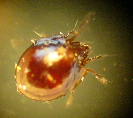
After some time, I managed to capture this tiny creature (the inner chamber of the cell is only a few mm across, so this is only about 0.5mm long) and took some pictures. It turns out that unlike many invertebrates, it is unable to walk on glass, so although its limbs were moving, the organism itself stayed still on a slide...
The tiny beast in question - a mite, possibly an oribatid. Note the shiny round carapace, bristly appendages and rostrum with transverse wrinkles.

The dorsal surface of the mite showing the even sculpturing.

The best close-up I could get of the head and front appendages/bristles.
So, as always when I decide to investigate a gall in detail, I have come away finding more than expected - it has induced me to read up on the unusual bacterium-mediated reporductive strategy of D. rosae, scrutinise the fine structure of something that is familiar on a macro scale only, and find an invertebrate that I genuinely can't identify (I await a friendly acarologist - should one appear, I'll post any updates). However, with its shiny single carapace, it does look like an oribatid (moss mite, order Oribatida). These vary in their diet, but different species feed on dead plant matter, fungi, carrion or lichens, while some are predatory. Given the microhabitat here, I suspect fungal and/or lichen feeding, and after a further search of the gall cells I found four of these mites. The gall and mites are now in a tiny vivarium, so I may get to see more behaviour and maybe young. If so, pics and details will of course appear here. Thanks for reading!


References

Csoka, G., Mattson, W.J., Stone, G.N. & Price, P.W. (eds) (1998). The Biology of Gall-Inducing Arthropods. General Technical Report NC-199, Forest Service, North Central Research Station, USDA, St Paul, MN. Contains many useful papers and used as a general reference in the publication below.

Redfern, M. (2011). Plant Galls. Collins, London. The source of much of the gall biology here, and a must for gall-nerds!

Wednesday, 25 May 2011

Galls of unknown origin

Those of you familiar with my blog will have noticed a minor fascination with galls lately, in part due to the publication of Redfern (2011). Following a couple of posts about the common Knopper Gall Andricus quercuscalicis, I felt it was time to tackle something distinctly less well known, to be precise, an undescribed species of the gall midge genus Dasineura (Diptera: Cecidomyiidae).

Dasineura ulmaria is a common galler of meadowsweet Filipendula ulmaria leaves, forming reddish-pink pouches with a hairy opening on the underside. However, in recent years (and indeed before, though less commonly), similar galls have been found on dropwort Filipendula vulgaris but with projections and openings on the upper sides of the leaves (see Harris 2010 for details of material seen between 2002 & 2009). As described in Redfern (2011, pp. 147-150), the newly hatched larva of D. ulmaria settles on a young leaf, and as it feeds, nutritive tissue develops beneath and around it, with cells growing and dividing to envelop it. This is typical of much insect-mediated gall formation where larval feeding induces gall production, although the precise mechanisms are porrly understood.With D. ulmaria eggs being laid on the underside of the leaf, the gall also has its opening here, unlike the galls on F. vulgaris as noted above.

Dasineura galls on F. vulgaris
These galls are single-chambered ('unilocular' in gall-speak) and as shown in the photo above, there may be more than one per leaf, and many per plant.

Dasineura gall on a single leaf of F. vulgaris. Note the hole towards the left which may be generalised plant-feeding damage or could be a potential predator of the enclosed larva.
Upper side of a F. vulgaris leaf showing two gall projections and openings.

Close-up of the gall openings showing fringing hairs and elongated cells of the conical projections.
Further investigation of the gall surface shows enlargement of plant cells, especially along the leaf ribs.

Enlarged cells on the outside of the gall, highlighted by reddening/darkening along ribs and veins.
Moving on from surface anatomy of the gall, its dissection reveals the single larva that lives inside.

The tiny (approx. 1mm long) larva in situ showing its yellow colour and segmentation. Also note the somewhat spongy texture of the nutritive cells lining the inside of the gall.

Close-up of the larva (width of photo approx. 0.5mm) showing the small head and tiny (paired) antennae. Also note the segmentation highlighted by dorsal and ventral sclerites, plus a hint of internal structure and gut contents.
In Dasineura ulmaria (and many other galling species), the young larva induces the production of large nutritive cells by the plant which line the gall chamber and provide it with food, especially during later stages/instars. As the larva grows, nutritive cells are consumed and nutrients continue to be supplied from vascular strands and stored starch. As the larva empties successive layers of nutritive cells, replacement nutrients have to pass across several dead layers. In D. ulmaria, nutrients also have to diffuse from vascular strands to the larva via the sclerenchyma - cells which have become somewhat woody but remain alive and connected - and it is possible that this is also the case with the galls on F. vulgaris. The larva pictured above is of the general form expected of Dasineura and although the papillae and structures known as the sternal 'spatula' (underneath the head end) which can be diagnostic (e.g. Smith 1989) were not visible in this young specimen, it is hoped that more mature larvae will be collected during a field visit in June to the same chalk grassland site that produced the specimen photographed here.

As noted by Harris (2010), this gall has been described previously - as D. ulmaria by some authors and D. harrisoni by others, although it may of course turn out to be genuinely undescribed. Therefore, until further work can be undertaken (e.g. on the host range of D. ulmaria and the identity of D. harrisoni which is unclear), this gall is to be treated as distinct but undescribed i.e. Dasineura (undescribed sp. A on F. vulgaris) - evidence that even in over-populated (and entomologist-laden) southern England, new species await discovery!

As ever, watch this space - if there are further developments I hope to be able to post them here.

References

Harris, K. (2010). Notes on gall midge galls recorded on Filipendula ulmaria and F. vulgaris in the United Kingdom. Cecidology 25(1): 6-10.
Redfern, M. (2011). Plant Galls. Collins, London.
Smith, K.G.V. (1989). An introduction to the immature stages of British flies. RES Handbooks for the Identification of British Insects 10(14):1-280.