Welcome

Welcome to my blog

This is where I post various musings about wildlife and ecology, observations of interesting species (often invertebrates)
and bits of research that grab my attention. As well as blogging, I undertake professional ecological & wildlife surveys
covering invertebrates, plants, birds, reptiles, amphibians and some mammals, plus habitat assessment and management
advice
. I don't work on planning applications/for developers. The pages on the right will tell you more about my work,
main interests and key projects, and you can follow my academic work here.
Showing posts with label bacteria. Show all posts

Wednesday, 28 March 2012

Keep feeling fasciation

Apologies for the terrible 1980s music pun - if you don't know what I'm talking about, Wikipedia will explain... Anyhow, today's topic is fasciation - the flattened expansion of plant parts into flattened bands or ribbons, sometimes with multiplication of flower heads (the phenomenon is also known as 'cresting'). Although generally considered rare as a whole, fasciation has been documented in well over 100 plant species in 107 families (Presland et al., 2009), including some very common ones such as the dandelion - often called Taraxacum officinale agg., but in the UK this is incorrect as T. officinale does not occur and there are around 235 species with intermediates and much variability. If you want to investigate this tricky taxonomic area in more detail, see Dudman & Richards (1997).

A common species of dandelion (Taraxacum sp., probably T. subhamatum) showing a fasciated stem with normal stems in the background.
Fasciation is not well understood in all cases, and can be caused by a number of factors - mutation of meristematic cells (i.e. abnormality of the growing tip), bacterial infection (particularly by Rhodococcus fascians), attack by mites or insect attack, or chemical, frost and/or mechanical damage - and in some cases can be inherited. In most cases, a single stem is affected (as in the photo above) and does not recur the following year.

Dandelion showing a fasciated flower-head.
One area where active research has explained fasciation well is the action of the bacterium R. fascians. Infection leads to hyperdosage of plant hormones, especially auxins and cytokinins which it may induce or may produce itself (Vandeputte et al., 2005). Induced overexpression of plant hormones can be complex, involving biochemical/transcription pathways which include genes and their homologues, hormone-inactivating compounds, hormone precursors, cofactors in various aspects of mineral metabolism and so on (Simon-Mateo et al., 2006). The visible effect of this occur by apical dominance being broken and secondary or auxiliary meristems being activated (hence the proliferation of flowers in the second photo).

Other causes are less well understood and may overlap with gall-causing agents in the case of mites and insects, while R. fascians is itself known as a causer of 'leafy gall' as well as fasciation (Redfern & Shirley, 2011). So, a feature to look out for, and one with plenty of opportunities for research - now, where's that funding application..?

References

Dudman, A.A. & Richards, A.J. (1997). Dandelions of Great Britain and Ireland. BSBI, London.
Presland, J., Oliver, J. & Barber, M. (2009). Abnormalities in Plants. Wiltshire Botanical Society.
Redfern, M. & Shirley, P. (2011). British Plant Galls (2nd ed.). FSC, Shrewsbury.
Simon-Mateo, C., Depuydt, S., de Oliveira Manes, C.L., Cnudde, F., Holsters, M., Goethals, K. & Vereeke, D. (2006). The phytopathogen Rhodococcus fascians breaks apical dominance and activate auxiliary meristems by inducing plant genes involved in hormones metabolism. Molecular Plant Pathology 7(2): 103–12.
Vandeputte, O., Oden, S. & Mol, A. (2005). Biosynthesis of auxin by the gram-positive phytopathogen Rhodococcus fascians is controlled by compounds specific to infected plant tissues. Appl. Environ. Microbiol. 71(3): 1169–77.

Thursday, 23 February 2012

Wheels of life

Straight in with a question today - have wheels evolved in nature? Now, I know it's been written about before, and there's no shortage of discussions on any number of online forums (or fora if you prefer), but it's something I've been musing on and coming up with some underlying questions - so, here goes with one my rare forays into the more speculative realms of biology and ecology...

Firstly, why might wheels be a useful adaptation? Well, they could provide an efficiency and simplicity of motion in some circumstances - I can certainly imagine animals using wide wheels to trundle across the soft sediments of the ocean floor for example (much like the wire-wheeled lunar rovers from Apollos 15-17). However, legs and fins generally work pretty well, with wheels really coming into their own on straight, smooth, hard surfaces. These are not common in nature, though humans produce plenty of them - and hence plenty of wheels. So, a lack of evolutionary advantage might be one reason why natural wheels are not widely seen.

Secondly, what do I even mean by a wheel? Here, I am only considering something that has an axle or bearing. There are plenty of organisms that roll - the South American pebble toad Oreophrynella nigra that tumbles down slopes to avoid predation, the wide variety of tumbleweed plants (and the rarer 'tumblefruits' such as Physaria) that disperse seeds as they roll with the wind, and the puffballs of the genus Bovista that are also blown around and so disperse their spores more widely. Ocean currents roll the coral Porites lutea across the sea floor and the small stomatopod mantis shrimp Nannosquilla decemspinosa can curl up and roll slowly like a wheel if stranded on a shallow damp sandy shore, thus returning to the sea. These are all interesting in their own right, and there are other examples, but none of them are wheels.

In fact, there don't appear to be any organisms that roll along on wheels in the way that humans' various vehicles do. As mentioned above, there may simply be no evolutionary pressure to produce a wheel, but there are also developmental constraints. For example, to have a wheel in a multicellular organism is tricky because, to be able to rotate freely, the wheel needs to be detached from the rest of the organism. If this is the case, how could it maintain a blood supply, neural connections and so on? Two options come to mind:

1. The wheel could be made of 'dead' material secreted by the organism, such as carapace material. This could grow as a toroid (doughnut-shaped) swelling on a limb/axle and gradually separate by thinning near the limb. This could produce a passive wheel on an axle much like a wood-turner produces a freely movable (but not removable) ring from a single piece of wood.
2. The wheel could be alive but self-contained. If a ring of cells developed as above and then detached, to be an effectively autonomous wheel, it would have to have its own energy supply (photosynthesis, chemosynthesis?) and so on.

Neither of these options have been discovered in nature, though this does not mean they never will - my feeling is that the lack of need is more likely to prevent wheels evolving than developmental problems. So far, I have not differentiated between passive and active wheels i.e. whether they simply roll like a cart (reducing the friction that would be caused by dragging) or are actively rotated by an energy source. Active wheels are developmentally even more problematic as a torque needs to be applied - in animals, motive force is produced by muscles, but this would not work on wheels as they need to be freely rotating. However, in bacteria, the problems of producing motive force, overcoming inertia and so on have been solved. In fact, the only example discovered so far of a true biological wheel (an active one that produces continuous propulsive torque around a fixed structure), is the bacterial flagellum, the  a propeller-like thread used for locomotion. Where the flagellum enters the cell membrane, there is a motor protein that works like a rotary engine, powered the flow of hydrogen ions (i.e. protons) across the bacterial cell membrane down a concentration gradient created by a proton pump. A similar system using a sodium ion pump exists in the genus Vibrio.

The structure of the flagellar base showing cutaway details of the 'motor'. Thanks go to Mariana Ruiz Villarreal for putting this and other diagrams in the public domain.

At an even smaller scale, the enzyme ATP synthase (which is involved in energy storage and transfer within cells) is somewhat similar to bacterial flagellar motors and is likely to be an example of modular evolution i.e. where two separate structures or sub-units (which evolved and previously functioned separately) become joined or associated, and in doing so gain a new function.

So, although true wheels have not been discovered in multicellular organisms, and both developmental and utility constraints make their evolution highly unlikely, maybe impossible, there are ways that wheels might be used in nature:

1. Through symbiosis, joining two otherwise unrelated structures/organisms in order to get round the developmental problems preventing direct evolution of wheels. This could be instinctive (imagine an extension of dung-ball rolling by dung-beetles) and is an idea which has been explored in fiction, e.g. in the Amber Spyglass (Philip Pullman, 2000). In this book, an alien race known as the Mulefa use large, round seed pods as wheels. They put these on sideways-oriented claws (which act as axles) on two of their legs, using the other two legs to push themselves along. The symbiotic aspect occurs because the trees that produce the seed pods depend on the rolling action under the weight of the Mulefa to break open the pods and allow the seeds to disperse and germinate. A number of other science fiction novels consider biological wheel use in a variety of ways, but Pullman's is probably my favourite so far, though other examples include David Brin's Brightness Reef (1995) and Infinity's Shore (1996), and Wheelers (2000), co-authored by Ian Stewart and Jack Cohen (who happen to be a couple of Terry Pratchett's collaborators if you like a bit of nerd-trivia).
2. Through tool use. Humans do this, using wheels widely - could other species do the same, even if with less technological sophistication? I'm just waiting to see corvids start rolling past...

OK, I think that's enough speculation for one day - if anyone out there does know of other examples of 'bio-wheels', I love to hear about them, so feel free to add a comment.

Wednesday, 15 February 2012

OMG in the OMZ: massive marine microbes

Today, I'm drawing inspiration from the Census of Marine Life, a decade-long project which has produced a huge inventory of marine life - a baseline catalogue to be used for further research and to inform the management and conservation of marine life. The Census looked at all scales from microbes to whales, at all latitudes and at all depths. The Census has produced a range of books, both popular and technical - one of the most straightforward and non-specialist, 'Citizens of the Sea' (Knowlton 2010) provided a couple of snippets that induced me to delve into the detail rather more...

First up, megabacteria - not the disease of budgies (which is actually a yeast), but very large true bacteria discovered off the coast of Chile and Peru in the 1960s. Placed in the genus Thioploca, the bacteria are filamentous and 2 to 7 cm (yes, cm) long. Secreting mucus, they form vast mats (the largest covering 130,000 km-sq) in/under the 'oxygen minimum zone' (OMZ), an area at 40-280 m with very little dissolved oxygen; instead they have to rely on hydrogen sulphide in the sediments. They oxidise this using nitrates (from sea water) which they can concentrate up to 500 mM in the liquid vacuole that occupies over 80% of their cell volume, even though the concentration of nitrates in sediment is only around 25 μM. Mucus-sheathed transport filaments send this nitrate 5–10 cm down into the sediment and reduce it, thus oxidising the hydrogen sulphide and creating a coupling of the nitrogen and sulphur cycles in the sediment (Fossing et al. 1994), producing pyrite and elemental sulphur as a result (Ferdelman et al. 1997). Thus, organic matter (in the form of anaerobic dissolved organic carbon) can be oxidised at low oxygen concentrations. The mats also provide food and shelter for a range of animals including squat lobsters (Pleuroncodes monodon), amphipods, and ophiuroids (Grupe 2011). As the OMZ shares features with conditions during the Proterozoic period (2.5 bya to 650 mya), and similar microfossils have been found, such bacteria may provide an insight into ancient life forms and ecology as well as performing a still little-known but key function in nutrient cycling. Research is ongoing with one recent example investigating Thioploca found in Danish waters where (in the species T. ingrica) nitrate accumulation was lower at around 3 mM, with bicarbonates and acetates used as carbon sources, and no mat being formed (Høgslund et al. 2010). 


A core from a Thioploca bacterial mat. The core is about 8 cm across and the mat about 1 cm thick. The mat is made up of many bacterial filaments with individual cells visible to the naked eye as white threads. Huge for bacteria! Photo courtesy of NOAA/Lisa Levin.
Now, ocean acidification due to carbon emission from fossil fuels may affect marine microbes - with microbial ecosystems responsible for between 50 and 90% of all marine biomass and over 95% of marine respiration, they maintain Earth's habitability though their influences on climate (they can sequester atmospheric carbon dioxide), nutrient cycling and the decomposition of pollutants (Leahy 2012). So, this could be very serious indeed and current research is looking at the  sensitivity of marine microbes to acidification. If I find links to results from this research, I'll post an update, plus I have some more bacterial and marine posts (among others) in the pipeline.

References

Ferdelman, T.G., Lee, C., Pantoja, S., Harder, J., Bebout, B.M. & Fossing, H. (1997). Sulfate reduction and methanogenesis in a Thioploca-dominated sediment off the coast of Chile. Geochimica et Cosmochimica Acta 61(5): 3065-3079. Fossing, H, Gallardo, V.A., Jørgensen, B.B., Hüttel, M., Nielsen, L.P., Schulz, H., Canfield, D.E., Forster, S., Glud, R.N., Gundersen, J.K., Küver, J., Ramsing, N.B., Teske, A., Thamdrup, B. & Ulloa, O. (1994). Concentration and transport of nitrate by the mat-forming sulphur bacterium Thioploca. Nature 374: 713-715.
Grupe, B. (2011). Sea Floor Habitats of the Chile Margin. NOAA Ocean Explorer [accessed 15/02/2012].
Høgslund, S., Nielsen, J.L. & Nielsen, L.P. (2010). Distribution, ecology and molecular identification of Thioploca from Danish brackish water sediments. FEMS Microbiology Ecology 73(1): 110-120.
Knowlton, N. (2010). Citizens of the Sea: Wondrous Creatures from the Census of Marine Life. National Geographic, Washington DC.
Leahy, S. (2012). Giant Bacteria Colonize the Oceans. Tierramérica. [accessed 15/02/2012].

Tuesday, 21 June 2011

What's in a gall? Part 3: Secrets of the bedeguar

About a week ago, I was on top of St. Catherine's Hill nature reserve near Winchester (Hampshire, England), leading a wildlife walk entitled 'Galls and other wildlife' as the heavens opened and the rain came down... and down... However, although few invertebrates were visible, there was one aspect of entomology that was visible whatever the weather - galls. Last month, I wrote about an undescribed gall species which can be found on this chalk grassland reserve, and before that I investigated the complex inner workings and inhabitants of a common gall species, the Knopper. So, when I saw a cluster of old woody bedeguar galls (Diplolepis rosae) on a sweet-briar (Rosa rubiginosa), I had to collect one and bring it back for closer examination.

A live gall of D. rosae


Bedeguar galls (also known as Robin's Pincushion in Britain) are the galls of the cynipid wasp D. rosae and are quite familar due to their large (a few cm across) spiky shape as shown above. Bedeguar is a Persian word relating to thistles (either their spininess or being wind-blown like thistledown), and there is evidence that ancient writers such as Pliny were familiar with these structures.

Most (well over 90%) of D. rosae wasps are female and it may well be that males are redundant and disappearing. As described in, for example, Csoka et al. (1998), this appears to be due to infection by the bacterium Wolbachia which causes reproduction to occur via thelytokous parthenogenesis (i.e. production of females from unfertilised eggs). Wolbachia has various effects on its different hosts (other wasps, woodlice, gnats, fruit flies) and comes in different strains, but it can interefere with meiosis (meaning females could not produce haploid eggs), cause sexual incompatibility, prevent production of males, or even feminise males (this happens in woodlouse hosts even though they have males genes). In any case, females emerge during May and June and lay eggs in leaf buds which are beginning to swell - whatever the precise criteria for bud selection, females can investigate a bud for up to an hour before deciding on its suitability. Inserting the ovipositir under a bud scale, eggs are laid between the developing leaflets inside  without damaging the plant tissues, and 30+ eggs can be laid, each in an individual cell.

As is usual in cynipid gallers, the leaflet cells around and below the egg immediately become highly active, enlarging and producing RNA, proteins and other substances, and a small pad is formed after about two days. Cell walls break down forming a cavity in the pad, and around a week after the egg was laid, it hatches and the larva enters the cavity. As it begins to feed, nutritive cells develop near its head - these line the chamber while the outside produces cambium. The epidermis meanwhile grows bulges which develop into the familiar multicellular hairs forming the outside of the gall which is familiar to us, the whole mass containing a number of cells. Also, new vascular tissues grow inwards to supply the nutritive layer and out into the hairs as well as linking with leaf veins. The gall is fully developed by July or August and at this point the larvae feed rapidly, being fully fed by October. They overwinter in the gall, pupate there in late spring, and new adults use their jaws to tunnel out (Redfern 2011).

An old bedeguar gall c. 25mm across showing exit holes in individual cells. The gall is also covered with lichens and moss.

The gall broken open to show vascular strands and the inside of a single cell.
The pictures above, especially the lower one, show the complex structure of the vascular tissues, shown here as spaghetti-like threads having grown through the outer layers before they became woody. As shown by the colonising lichens etc., this is quite an old gall and so no D. rosae or associated parasites/inquilines are present, but the complex structure is of interest, especially given the possibility of non-galling invertebrate colonists. A variety of structures is shown in the following pictures:

Enlarged cells similar to those seen in the Knopper gall (see link at top of article)

Layering of cells in the bedeguar gall.

Spongy texture of woody cells surrounding a gall cell.

Some of the vascular tissues, now woody, linked to various parts of the gall and host plant, including the hairs - elongate plant cells are visible.

A section from around a gall cell showing the same elongate structures and their association with the spongy layer.
Looking inside an individual gall cell, it is clear that there is colonisation by, for example, lichens and fungi, but there is evidence of other biota using these ready-made structures.

The inside of a gall cell showing a white membrane suggesting a cocoon, plus black specks of 'frass' (invertebrate faeces).
Looking behind this membrane, among the green algae and black frass, there appears to be an empty skin (exuvium) - the small linear structures in the centre are probably legs.
Having found an exuvium, I had to wonder what had left it there. I didn't expect to find anything, but then there was a tiny flash of reddish movement as I looked down the microscope.
In the bottom of the open cell, a small, round (and quickly moving) red shape.
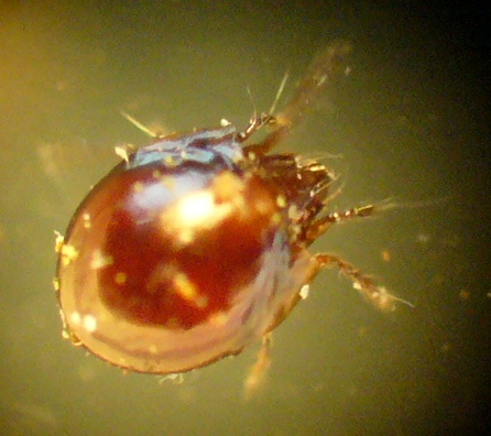
After some time, I managed to capture this tiny creature (the inner chamber of the cell is only a few mm across, so this is only about 0.5mm long) and took some pictures. It turns out that unlike many invertebrates, it is unable to walk on glass, so although its limbs were moving, the organism itself stayed still on a slide...
The tiny beast in question - a mite, possibly an oribatid. Note the shiny round carapace, bristly appendages and rostrum with transverse wrinkles.

The dorsal surface of the mite showing the even sculpturing.

The best close-up I could get of the head and front appendages/bristles.
So, as always when I decide to investigate a gall in detail, I have come away finding more than expected - it has induced me to read up on the unusual bacterium-mediated reporductive strategy of D. rosae, scrutinise the fine structure of something that is familiar on a macro scale only, and find an invertebrate that I genuinely can't identify (I await a friendly acarologist - should one appear, I'll post any updates). However, with its shiny single carapace, it does look like an oribatid (moss mite, order Oribatida). These vary in their diet, but different species feed on dead plant matter, fungi, carrion or lichens, while some are predatory. Given the microhabitat here, I suspect fungal and/or lichen feeding, and after a further search of the gall cells I found four of these mites. The gall and mites are now in a tiny vivarium, so I may get to see more behaviour and maybe young. If so, pics and details will of course appear here. Thanks for reading!


References

Csoka, G., Mattson, W.J., Stone, G.N. & Price, P.W. (eds) (1998). The Biology of Gall-Inducing Arthropods. General Technical Report NC-199, Forest Service, North Central Research Station, USDA, St Paul, MN. Contains many useful papers and used as a general reference in the publication below.

Redfern, M. (2011). Plant Galls. Collins, London. The source of much of the gall biology here, and a must for gall-nerds!

Saturday, 19 February 2011

Entomology of Star Wars. Episode II: Metallic snails and vent mussels

As Episode I proved so popular, I've been thinking about extending the 'Entomology of Star Wars' into an occasional series - not easy as I don't go in for a lot of 'speculative biology', but I'm up for a challenge and do have a penchant for Science Fiction. So, time for a bit of a nerd-fest...

One beast that I have wondered about is the big asteroid-dwelling worm that the Millenium Falcon encounters in 'The Empire Strikes Back' - what it's made of, what it lives on, how it survives in space and so on. A little online digging soon told me that it's called a 'space slug' or 'exogorth' and that it's a silicon-based gastropod (I'm pleading poetic license and sticking with 'entomology' rather than 'conchology' though), feeding mainly by metabolising asteroid minerals through its root-like tail and absorbing stellar energy, though not averse to eating the occasional spacecraft or other unwary space-dwelling creatures. Also, it apparently reproduces by fission, moulting as it grows, then simply splitting in two when large enough.

The exogorth - in space, no-one can hear you roar!
Now, I don't intend to get into the whole hypothetical silicon-based life/alternative biochemistry thing - there are plenty of people doing that already, and there's even a Wikipedia page about it here. Instead I'd like to see if there are any parallels between this fictional beastie and real-world organisms.

Firstly, the metabolising of rocks and their constituent minerals. It's well known that some bacteria (lithoautotrophs) can metabolise a variety of minerals such as sulphur, iron and manganese and are involved in both the creation of limestone cave systems (speleogenesis), and the phenomenon of acid mine drainage (AMD), the outflow of acidic water from metal and coal mines. The biochemistry of such acidophilic bacteria can be complex, but in the case of cave systems such as Carlsbad and Lechuguilla in the US, rocks have been attacked microbially in three ways:
  • Oil-metabolisers - their biochemistry produces hydrogen sulphide which in turn produces sulphuric acid.
  • Rock-eating bacteria - the lithoautotrophs directly metabolising minerals.
  • 'Snotites' - large bacterial colonies which are primary producers in such ecosystems and drip sulphuric acid.
So, maybe the exogorth has lithoautotrophic gut flora - it has a mouth and dentition, so a gut seems likely. Thinking of a worm-like creature with symbiotic bactera does lead neatly onto the annelid tube worms such as Riftia associated with hydrothermal vents. These have become familiar creatures over the last decade or so through various TV programmes, and although they do not have a mouth or gut, they do contain symbiotic bacteria. These live within a specialised organ (the trophosome) in the worm and metabolise compounds such as carbon dioxide and hydrogen sulphide which are absorbed by the worm's plume. In turn they provide carbon compounds which nourish the worms. Similarly - and providing a handy molluscan parallel with the exogorth - vent mussels (Bathymodiolus thermophilus) are almost entirely dependent on symbiotic bacteria in their gills. Not only that, but in 2001, the Scaly-foot Gastropod (Crysomallon squamiferum) was discovered associated with hydrothermal vents in the Indian Ocean. What is unusual about this species is that its foot is armoured with 'scales' (technically 'sclerites') of iron sulphides (greigite and pyrite) and that its shell has a third layer also containing iron sulphide - it is the only animal known to use this mineral skeletally in this way. This means we do have molluscs with mineral-metabolising symbiotic bacteria, and that at least one can incorporate what we consider 'unusual' compounds into its anatomy. But, what about reproduction?

A heap of chemosynthetic vent mussels.
This seems fairly straighforward - all molluscs reproduce sexually (most gastropods are hermaphroditic), so definitely no fission. Still, although asexual reproduction is rare in higher animals, they are not unknown. For example, in Mexico, there are Topminnows of the genus Poeciliopsis where it is seen. Two species, P. monacha and P. lucida, reproduce sexually with their own species members when separate, but where they co-exist, they hybridise to produce all-female forms that reproduce by cloning (hybridogenesis and gynogenesis). In hybridogenesis, the female mates with a male, producing female offspring with both the maternal and paternal genomes. When that female produces eggs, the male genome is discarded, leading to the all-female form. In gynogenesis, females are triploid and also mate with males, but the male genome does not contribute to the offspring. There's also a good diagram explaining the process of hybridogenesis in water frogs here and there's plenty of research into gynogenesis in the African Clawed Frog (Xenopus laevis) - certainly, amphibians do seem to have a flexible approach to reproduction...

So, parallels between exogorths and real-world organisms? For molluscs, nutritionally yes but reproductively no - but what about the whole 'surviving in a vacuum' issue? Well, I think I'll leave that for Episode III...