Welcome

Welcome to my blog

This is where I post various musings about wildlife and ecology, observations of interesting species (often invertebrates)
and bits of research that grab my attention. As well as blogging, I undertake professional ecological & wildlife surveys
covering invertebrates, plants, birds, reptiles, amphibians and some mammals, plus habitat assessment and management
advice
. I don't work on planning applications/for developers. The pages on the right will tell you more about my work,
main interests and key projects, and you can follow my academic work here.
Showing posts with label pupae. Show all posts

Friday, 20 March 2015

Unexpected egg-flies

Back in January, my wife showed me a blackbird egg she'd found in the garden. It looked whole, but was incredibly fragile and broke almost as soon as I picked it up, clearly already being cracked. I expected maybe a whiff of something nasty, but there was more going on than I expected...

Inside the egg... lots of invertebrate action.
Naturally, instead of saying 'yuk' and throwing it away, I had a closer look. The orange inveretebrates are mostly fly pupae plus a few larvae. The black shapes are pupal skins of something already emerged, and the large larva in the middle something else entirely. A quick look in Smith (1989) jogged my memory that the smaller ones were probably juveniles of the Psychodidae, also known as moth-flies or owl-midges. The larvae of many species are associated with decaying organic matter, so the inside of a failed egg is a plausible hiding/feeding place whether the goo insode is egg material or something else that had seeped in. Beyond that I couldn't tell, so I put them in a hatchery and waited. After not too many days this is what I found...

Pupae, pupal cases and adults of a psychodid fly.
In total there were 15 pupae and these soon started to emerge as adults. The small size, their shape, and their hairy/feathery wings mean they don't look like typical flies, but that is exactly what they are - as you can see below, they have a single pair of wings, the other pair reduced to drumstick-shaped balance-organs (halteres). Psychodidae are not an easy group to work on (especially if you are not that familiar with them), and identifiying the flies to genus, let alone species, would require time, a microscope, and a copy of Withers (1989). I won't go through all the steps, but here are a few key features:

There are two veins (dashed lines) between the two main forks in the wing veins (circled).
The arrow indicates the eye-bar - an extension of the eye above the antenna. The circle indicates the pale haltere mentioned earlier - yes, these realy are flies!
 The antennae are an important feature too, but take care when keying out genera and species...

The key asks whether the antennal segments are barrel-shaped or have an elongated stalk. These look barrel-shaped but...

...if you remove a segment you can see that the barrel-shape in this case is formed of hairs that hide the real shape - the dark segment clearly has a stalk.
So, after quite a bit of deliberation, this keyed out as the genus Psychoda. I'm less certain about the species (not only can they be difficult to separate, but the taxonomy of Psychoda needs to be revised), but it might be P. alternata (the stripy larva in the first photo matches this, as does the foul habitat). Withers (1989) doesn't list Hampshire for this species, but the Psychodidae are under-recorded, and he does mentioned Wiltshire and Sussex, so it wouldn't be a surprise. That's enough for now - I shall leave you with some more juveniles, feeding/hiding merrily in the goo...

Psychoda - three pupae (complete with a pair of pupal horns for breathing) and a larva.

References

Smith, K.G.V. (1989). An Introduction to the Immature Stages of British Flies. Handbooks for the Identification of British Insects 10(14): 1-280.
Withers, P. (1989). Moth Flies. Diptera: Psychodiae. Dipterists Digest 4: 1-83.

Monday, 28 July 2014

Nest of tiny delights

Wow - a month since my last post - there has been no shortage of wildlife to write about, just no 'spare' time to do the writing. However, the paid stuff is quiet for a few days at least, so I thought I'd share some observations of the small and hidden, to be precise what I found lurking within a used-and-the-young-fledged robin's nest.

The robin's nest before dismantling.
You never know what'll be hiding in a nest (whether bird or mammal) - there's always the possibility of some under-recorded parasites, plus the array of small invertebrates that simply use the structure as their own. So, a white tray, various tweezers and pins and some time later, this is what I found, along with quite a few Collembola (springtails) and Psocoptera (barklice) which I didn't collect on this occasion as I knew I would have time to work through the identification.

One of three similar pupae a few mm long, currently in my hatchery.
Another pupa, also a few mm long.
A very young spiderling, highly active and I think one of the wolf spiders (Lycosidae)
A tiny dead wasp, probably one of the Pteromalidae, possibly predated by a spider.
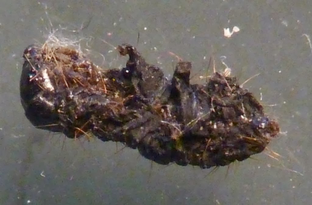
The exuvia (skin) of, well, I'm not sure...

Alive and active, the larva of what is probably a 'carpet beetle' (Dermestidae).
So, not much in the way of definite identifications - with juvenile stages, that tends to be difficult, though if any are successfully raised to adulthood, I will as ever update the post. Also, no parasites, but there was good evidence of a thriving community of small invertebrates potentially living out their whole life cycle in the structure of the nest with larvae, pupae, evidence of emergence, predators and prey.

Friday, 28 March 2014

After the wet (part 2)

Having recently raised a Bright-line Brown-eye moth Lacanobia oleracea from a pupa rescued from our storm-damaged fence panels, this morning I awoke to find another new arrival in the hatchery, a Brimstone moth Opisthograptis luteolata - not to be confused with the Brimstone butterfly Gonepteryx rhamni.

Brimstone moth Opisthograptis luteolata.
This species feeds on a range of shrubs and small trees - although none of its more common host plants grow in our garden, there are plenty nearby and the overlapping fence-panels must have provided effective overwintering shelter until it blew down. Adults are usually found from April to October so this one was only a few days early.

Portrait of the Brimstone moth Opisthograptis luteolata.
Empty cocoon and pupal case of Opisthograptis luteolata.The pupa was inside the cocoon which is usually a rounded elliptical/oval shape - here it is squared off and has flat sides due to being formed between flat fence panels.

Monday, 17 March 2014

After the wet

It seems that our several months of almost incessant rain has finally come to an end, and spring is happening. Although I expect that some species will have suffered due to flooding (water vole burrows, soil-hibernating invertebrates...), some are starting to appear. So here's a quick non-technical introduction to a couple of garden-dwelling moths that have made themselves known to me recently.

A mature larva of the Old Lady moth Mormo maura. Note the bright orange spiracles along the side, and the black marks in the rear half. Approx 60mm long.
Head of larva of Mormo maura - note the shiny, speckled head capsule with ocelli (simple eyes) and small antennae
M. maura is a common species and I have previously seen adults in our garden, including one roosting in a shed. Ivy (Hedera helix) is one of its main food plants and we have plenty of that so it is not a surprise to see this species.

A couple of months ago, a series of storms destroyed many garden fences (not to mention causing widespread flooding) in the UK and while clearing up the debris, I found several pupae that had been dislodged. Most were put in our 'bug hotel' but a few were taken indoors to be hatched. One of these emerged a couple of days ago and proved to another common species, the Bright-line Brown-eye Lacanobia oleracea. It feeds on a wide range of woody and herbaceous plants and again is no surprise, but did provide some photo opportunities prior to release.

L. oleracea - the bright line is along the rear edge of the wings, the brown eye refers to the kidney-spots in this species.
Close-up of the wings showing the scale-patterns.
Side view showing the hairy and tufted thorax.
Portrait!

Wednesday, 5 February 2014

The Great Juvenile Chrysomelidae Project

You know I like invertebrates, especially insects, in particular beetles, and that I have an inordinate fondness for the Chrysomelidae (leaf beetles). You may even know that I published an identification key to the adults of British species. However, I have (foolishly?) decided that this isn't enough and am therefore embarking on a project to (eventually) produce some sort of identification guide to juvenile chrysomelids - eggs, larvae and pupae.

Why? Well, for many of the species, the ecology and life histories are poorly understood, and many juvenile stages are undescribed. So, it's first of all, an opportunity to summarise where more research/beetle-hunting is required. If the larva of species X has never been described, then there's an opportunity to try to find it. This allows the biology of the group to be better understood and in turn, conservation measures to be more appropriately designed and applied. The better we know them, the better we can protect them.

This is why the first step is the creation of a spreadsheet covering what is known/published for each species on the British list so that I can see where the genuine gaps in our knowledge lie. I have a draft of this spreadsheet and what I'm after is feedback to see what I've missed - presumably some stages/species are covered in publications, grey literature, theses etc that I've not managed to find, as well as observations that have not been written up in keys or formal descriptions. Some blanks could be filled in with a fair level of confidence e.g. more 'earthen cells' for pupal location, but I've erred on the side of caution and left blanks where there's no published (or directly observed) confirmation I know of. There's also a list of all the references I've found so far.

Interested? want to help?

If you'd like to check the spreadsheet and see if there's anything you could add to it, please get in touch so I can email it to you (if there was a wiki function here, I'd use that, but there isn't...). Then simply send any additions to me citing the cell number in the spreadsheet where it should go (either by email or a comment here).

I hope that all makes sense and look forward to seeing what I've missed - I suspect this is going to be a long project...

A chrysomelid larva, but which one?

Tuesday, 10 December 2013

A tiny clue that looks like poo

Sometimes a seemingly innocuous observation can lead onto an interesting ecological story... A couple of days ago while collecting bramble and raspberry leaves for my stick-insects, I noticed a small black-and-white cocoon attached to the underside of one of the leaves.

Black-and-white cocoon, 6mm long
Although the occupant has clearly emerged as an adult, I wondered if the distinctive black-and-white cocoon might be identifiable, at least to family level or similar. A quick web search made it clear that this was the pupal cocoon of an ichneumon and it didn't take long to find out that the pattern (which maybe camouflage it as bird-dropping or piece of mould?) is characteristic of the genus Hyposoter (subfamily Camploplaginae).

At least 15 species of this genus can be found in the UK and they parasitise the caterpillars of various butterflies and moths such as the Lymantriidae (tussock-moths), Pieridae (butterflies - whites and yellows) and Lycaenidae (butterflies - coppers, hairstreaks and blues). They do so by the female wasp laying an egg in the host caterpillar, piercing it with her ovipositor. Once hatched, the wasp larva develops inside its host which shrinks, becoming a hard,  brittle shell - effectively mummified. When ready to pupate, the wasp larva spins a cocoon inside the larval skin (or in some cases the host skin splits and the wasp pupates outside) and this creates the black-and-white patchwork pattern - it takes about a month from the egg being laid to a new adult emerging.

I don't know which Hyposoter this is and I doubt that it's possible to tell from just a cocoon. However, one of the British species (H. ebeninus) has been filmed going through its life cycle, and this is certainly a candidate for the species here as the adults match ichneumons I have seen but not identified (though that isn't a reliable indiactor - they are a diverse and tricky group to ID), and parasitises the cabbage white butterfly (Pieris brassicae) which is common here. Again, this is not enough for an identification, but it is something I can look out for next season. In any case, I hope that's an interesting little ecological tale from a passing observation.

Close-up of the Hyposoter cocoon showing silk threads attached to the host's mummified skin.

Monday, 18 November 2013

Multiple cocoon of many mysteries

I'm not often stuck for an identification, at least not for long but while dismantling an old fence panel a few days ago, I did find some things I really couldn't identify, at least maybe not...

Cocoons found attached to an old fence panel
The left-hand one is in some ways the easiest - it's probably a moth, maybe the knot grass (Acronicta rumicis) which I have seen in our garden and which makes long grass-covered cocoons (this one is around 55-60mm long including the grass) - hopefully I'll find out for sure when it emerges as it's now in one of my hatcheries.

The other one is less straightforward. As there's a cigar-shaped cocoon inside it, it is more of a nest than a cocoon as such, and appears to have two concentric 'walls', the inner one thicker and covered in tiny wood fragments.

The 'inner' nest around 18mm long, the cocoon inside is around 8mm long.
The cocoon within the inner nest - whatever was inside has clearly emerged, leaving an open end with a small 'lid'
Next to the cocoon, there's clearly a small black shape, plus in the very top photo, there's material between the two nest walls.

The small black shape turns out to be a dead and shrivelled caterpillar - whatever it was, it bore long hairs/bristles which can still be seen. Length approx. 6mm
From this angle, the true legs can be seen towards the head end (left) curled under the thorax, while the larger, stubby prolegs are more clearly visible in the middle/rear
The relatively simple lens/eye arrangment of the caterpillar
I can't tell what this caterpillar is as it is dry and shrivelled, but the form of the inner nest suggests a puss moth (Cerura vinula) or other member of the family Notodontidae. Mature puss moth larvae are large and spectacular, but the first instar is small and black. However, it doesn't have long bristles, so can't be this species, though other members of the family may be plausible candidates. A different question does arise though - cocoons give rise to adults, not larvae, so what is it doing here? If there were wasp eggs, then one plausible explanation would be that it was brought in immobilised by a solitary wasp as a food source for its own young. However, this is a nest with a cocoon in it, not eggs. Also, there is no evidence of a wasp in the cocoon, but there is the material between the inner and outer nest walls. I am unaware of any wasps where a host caterpillar is entombed by a pupating larva to await its adult emergence, but they are a diverse group  often with complex (and often poorly understood) life-cycles, so I imagine it is possible. Alternatively, the caterpillar was simply caught inside a nest construction and starved.

Spider exuviae (moulted skins) within the outer nest
As seen in the photo above, the material consists mainly of moulted spider skins (exuviae) of differing sizes, sugessting an individual spider grew and moulted several times using the thin outer nest (the red structure top-left is a set of mouthparts, almost as large as the whole skin bottom-right). It seems plausible that the hiding place was a good one and a spider simply used it, building its nest around the tougher one already there. Looking even closer there is more to see.

The thin outer wall and the thck inner wall with wood shavings. Just below where they meet, a small white structure is visible in partial shadow.
Hidden behind the larger structures, what appears to be another, much smaller cocoon. This is now also in a hatchery awaiting whatever emerges.
This looks like an opportunistic cocoon-builder that has just used a handy crevice, but it may be related to the larger nests. I don't know which species are involved, but I hope that the two remaining cocoons remain viable and I'll see which species emerge. You never know, it might shed some light on the mystery nest/larva - and if it does, I will, as ever, post it here.

Tuesday, 10 September 2013

Grubs within grubs within grubs

Back in the 17th century, Jonathan Swift (he of Gulliver's Travels fame) wrote:

"So nat'ralists observe, a flea
Hath smaller fleas that on him prey;
And these have smaller fleas to bite 'em.
And so proceeds Ad infinitum."

Then, in the 19th century, mathematician Augustus De Morgan updated this to the more familiar

"Great fleas have little fleas upon their backs to bite 'em,
And little fleas have lesser fleas, and so ad infinitum."

Well, this might not proceed quite ad infinitum, but there certainly can be several tiers of parasitism. One common example involves the small wasp Apanteles glomeratus (family Braconidae) which parasitises larvae of the 'cabbage' white butterflies Pieris brassicae and P. rapae. Eggs are laid into the butterfly larvae, where they hatch and develop - after about 15 to 20 days the wasp larvae emerge and the host dies. The wasps then pupate in silken cocoons next to the dead host, creating the arrangement that can be seen attached to a wide range of substrates, sometimes in very exposed situations (such as the middle of a glass patio door...), with the whole cluster attached by silk threads.

A typical arrangement of Apanteles glomeratus cocoons by the dead host larva of Pieris brassicae.
Apanteles glomeratus cocoons showing the tangle of silk threads.
I have seen  numerous examples of this arrangement over recent days, largely because following several very poor summers in the UK, Pieris spp. are some of the few butterfly species to have done well, and consequently there are many of their larvae for parasites to target. Also, as Pieris spp. are ravenous eaters of brassicas in farms and gardens, A. glomeratus is a vegetable-grower's friend. However, if I look more closely, I might find 'hyperparasites' (parasites of parasites) such as the tiny ichneumon wasp Lysibia nana which targets braconids such as A. glomeratus. Closer still and - well, I don't think anyone has investigated - but I'd expect to find microscopic protozoan, fungal or nematode parasites. Maybe I need to capture some L. nana and have a look.

If you would like more technical detail about parasitic wasps, Ronquist (1999) gives details of their phylogeny, classification and evolution (including, in Table 1, some examples of who parasitises who). In contrast, if you fancy an excellent (and silly) photo of a braconid larva, try here instead (or, as well - I like both, and Myrmecos is a fine blog looking at entomology, often ants, from a more photographic viewpoint).

Reference

Ronquist, F. (1999). Phylogeny, classification and evolution of the Cynipoidea. Zoologica Scripta 28(1-2): 139-164.

Wednesday, 26 June 2013

It's June, and wasps are busting out all over

Bees are good. We like bees. They pollinate and are pleasant to watch. They are of course also wild creatures subject to the stresses that any other organism has to deal with, and one of these is parasitism. With our garden bee logs, new and old, almost completely occupied by the red mason bee Osmia rufa, it is not surprising that other nesting sites are sought out. Sometimes the chosen spot is not a good one, as in this case where a nest had been attached between a hoe and the inside of our tool shed - we'd noticed the nest, but the hoe fell down anyway, taking some of the cells and cocoons with it.

Osmia rufa nest - the open sides of the cells were pressed against the handle of a hoe until it fell down. Note the larva in the bottom centre cell (now taken away to be raised indoors)
The inside of an O. rufa cell - note the plug made of numerous small pieces of mud tamped into place by the prongs on the female's head.
A cocoon and pollen ball (larval food supply which includes a small amount of nectar) from an O. rufa cell.
Being a fan of bees (well, wildlife in general really) and ever-curious, I collected the cocoons that had been displaced (about a third of the whole nest) and brought them indoors to be put into a hatchery. I was of course hoping to see the bees emerge and release them, but a few days later something else happened entirely; the hatchery was full of small parasitic wasps - Monodontomerus obscurus (Hymenoptera, family Torymidae) - 21 in total.

Some of the Monodontomerus obscurus hatched from O. rufa cocoons
A female (note the ovipositor) M. obscurus apparently feeding on an O. rufa larval pollen store.
Although the larger parasites of O. rufa are listed in Baldock (2008), there seems to be relatively little mention of this aspect of the bee's natural history even though it is a familiar and popular species. However, Krunic et al. (2005) provide more detail and note that M. obscurus can suppress O. rufa populations in some nesting situations such as paper tubes and hollow reeds.

This is a normal part of bee ecology of course - and the wasps are interesting little jewels in their own right, so (as ever) I took the opportunity to have a look at one of them (a female) more closely. Males are present but smaller (approx. 4mm long without appendages, females approx. 5mm) and lack the ovipositor that female use to lay eggs into the bee cocoons.

Female M. obscurus - approx 5mm long plus 2-3mm of ovipositor
Close-up of the head of a female M. obscurus - note the red eye with short white bristles, plus the sculpturing on the head and thorax.
Close-up of the hind femur of a female M. obscurus - note the sculpturing on the leg and the 'finger-print' pattern of sculpturing on the abdomen behind it. Also, the edges of the thoracic plates bear fringes of short white bristles, presumably to protect the membranous joints.

O. rufa cocoon with M. obscurus exit hole.
With the cocoon having disgorged its parasites, there was only one thing left to do - open it up to what was left inside.

The content of an O. rufa cocoon after emergence of M. obscurus.
I did wonder of there would be much evidence of the juvenile bee, but very little was left, having presumably been consumed by the wasp larvae - there are a few fragments (e.g. bottom right) but nothing more. Most of the invertebrate material consists of pupal skins of M. obscurus - unsurprising given the number that emerged. There is some frass (faeces) and bits of dried mud from the nest which must have fallen in before the cocoon was spun. Top left is a dead female wasp who didn't emerge fully from pupation. She is of course also in a hatchery just in case she releases smaller parasites of her own...


References

Baldock, D.W. (2008). Bees of Surrey. SWT, Woking.
Krunic, M., Stanisavljevic, L., Pinzauti, M. & Felicioli, A. (2005). The accompanying fauna of Osmia cornuta and Osmia rufa and effective measures of protection. Bulletin of Insectology 58(2): 141-152.

Tuesday, 30 April 2013

Tree-moth mystery solved

After much musing about whether I had found Stigmella suberivora on the non-native cork oak Quercus suber, a discussion on iSpot followed by a little extra microscope work shows it to be Phyllonorycter messaniella. The identification was finally confirmed by looking at the intersegmental spines which are particularly long in the pupa of this species.

The long intersegmental spines which are diagnostic for the pupa of Phyllonorycter messaniella.

This is a widespread species in the UK on evergreen oak Q. ilex and various deciduous oaks, as well as some other tree genera (Heath & Emmet, 1985). However neither this book nor the excellent leaf-miner websites here and here give Q. suber as a host for the moth in the UK. The next job is to find out whether it really is a new host for the moth in this country... update when I know more.

Reference

Heath, J. & Emmet, A.M. (eds.) (1985). The Moths and Butterflies of Great Britain and Ireland. Volume 2: Cossidae - Heliodinidae. Harley, Colchester.

More on the tree-moth combo

A few weeks ago, I posted about leaf-mines on cork oak (Quercus suber) and whether I had found the first British record of the moth Stigmella suberivora on this tree species (it is known from Q. ilex in Britain and Q. suber in continental Europe). Although the mine appeared correct, this is not enough to confirm the record, so I revisited the tree and collected a couple of leaves with occupied mines in order to, hopefully, raise adults which can be positively identified. This is a somewhat uncertain activity but the pupae I collected are being kept in suitably (but not too) moist/humid conditions and, as I write, are definitely still alive.

I have added a couple of images of the pupae, though I have noticed that whereas adults, and to a lesser extent eggs and larvae can be found figured in various publications, reference images of pupae are difficult-to-impossible to find, even on the excellent UK Moths site. So, here they are - fingers crossed that my next post on this topic will cover the final identification!

Unidentified pupa from a Q. suber leaf mine - approx 4mm long - note the mobile abdomen.
Unidentified pupa from a Q. suber leaf mine - head to the left, approx 4mm long, note ventral thoracic hooks and the wings dorsally (beneath in this photo).