Welcome

Welcome to my blog

This is where I post various musings about wildlife and ecology, observations of interesting species (often invertebrates)
and bits of research that grab my attention. As well as blogging, I undertake professional ecological & wildlife surveys
covering invertebrates, plants, birds, reptiles, amphibians and some mammals, plus habitat assessment and management
advice
. I don't work on planning applications/for developers. The pages on the right will tell you more about my work,
main interests and key projects, and you can follow my academic work here.
Showing posts with label algae. Show all posts

Tuesday, 19 April 2011

Diary of a farm pond: April 2011

As you may know from one or two other posts, and the 'Wildlife of Highbridge Farm' page over on the right, about a year ago I got involved with a community farm project at Highbridge Farm, Hampshire, southern England. Although its main function is to grow cheap, chemical-free food without reliance on oil/petroleum products (it's a project of the local transition group, ETNet ), this comes with an interest in, and reponsibility for, wildlife and the wider environment. So, formed from an old farm gravel pit, a pond has been created and I intend to follow its development as a wildlife habitat.

The pond/gravel pit, summer 2010
Early on in the project, the pond was essentially a gravel pit which had partly filled with water and become colonised by ruderal marginal plants plus dense floating algae. Obviously this has some wildlife benefit and a few common species of Odonata (dragonflies and damselflies) were seen using it, but the idea was to enhance its ecological value. The green tarpaulin covers a small water pump as the pond is used for irrigation when necessary - something that will continue as the habitat develops.

The pond, April 2011
Over the 2010/2011 winter, many of the ruderals were removed, along with the blanket of algae. The second photo shows that the sides have also been reprofiled, and on the right is an area which has been sown with native wild flower seeds suitable for a stony substrate - behind these (off-photo) are rows of native fruit and nut trees. Some of the ruderals remain or will be allowed to return - after all, nettles (Urtica) and docks (Rumex) are important for many invertebrates - but this work should provide greater biodiversity. However, there's only way to really see how effective such conservation work is, and that is to monitor what lives there. Sounds like an excuse to rootle about for invertebrates...

Although mid-April is a little early for Odonata, on 16th April there were two exuviae (skins) of nymphs that has crawled up marginal plants and emerged as adults.

An exuvium - but which species?
The relatively small size (approx 15mm long plus another 5mm or so for the caudal lamellae at the end of the abdomen which have stuck together) and narrow form make this a damselfly rather than a dragonfly, and so I headed for the excellent little book by Cham (2009). Given the early time of year, there aren't many possible species and I was guided by which I had seen last year. So, I soon came to the Large Red Damselfly (Pyrrhosoma nymphula). This species is relatively stout for a damselfly nymph/exuvium and matches the photo very closely - it has relatively long wing sheaths and a short abdomen, and the back of the head has a rectangular shape (ignoring the central dent).Before emergence the femora would have faint dark band and the lamellae would usually have pale markings but these have now disappeared. I haven't seen this year's adults yet, but at least I know they are probably around somewhere.

A variety of other invertebrates are present at the pond - whirligig beetles (Gyrinus sp.), pond-skaters (Gerris sp.), honey bees, some as-yet unidentified diving beetles, springtails (Collembola) and wolf-spiders (Lycosidae, especially Pardosa sp.) on marginal stones, and water snails; however it is the flies (Diptera) that currently appear to be most diverse (or at least most obvious).

The first recorded this year was a pair of craneflies Tipula vernalis, a spring species mating on a small remaining patch of floating algae in late March. Soon after in April, the bee-fly Bombylius major was noted, along with others including the non-biting midge Chironomus anthracinus, the hoverfly Rhingia campestris and the moth-fly Boreoclytocerus ocellaris though it's worth noting that some of these need collection and closer examination to identify them with certainty. Moth-flies are certainly under-recorded and it's worth getting a copy of Withers (1989) if you are interested; it's currently available for just £4.

Tipula vernalis, busy making more craneflies

Male (plumed antennae) Chironomus anthracinus

The moth-fly Boreoclytocerus ocellaris

The hoverfly Rhingia campestris showing the long rostrum
There's already more (with a few more pics below), but I think initial indications are good - a wide range of colonists already this year to supplement the species persisting from before conservation works on the pond. It is unclear what impact the current dry spring weather is having, though the pond is likely to have some hydrological connection with the nearby River Itchen so hopefully water levels won't get too low. Anyhow, further updates will follow - until then, enjoy the following:

A honey-bee Apis mellifera; a very orange form, maybe the Italian strain?

Tadpoles of common frog Rana temporaria, plus a water snail


References

Cham, S. (2009). Field Guide to the Larvae and Exuviae of British Dragonflies. Volume 2: Damselflies (Zygoptera). British Dragonfly Society, Peterborough.

Withers, P. (1989). Moth Flies. Diptera: Psychodidae. Dipterists Digest 4: 1-83.

Friday, 8 April 2011

What's in a gall? Parasites and photobionts...

Something that has begun to fascinate me over the last couple of years is cecidology - the study of galls. I am by no means an expert in this, though I have been drawn to joining the British Plant Gall Society (BPGS) and last year even led a gall-hunting trip (another one's booked in for later this year). Why? Well, good question - it's a combination of factors - the diversity of forms, the fact that finding and IDing galls is often easier than finding and IDing the tiny invertebrates that create them (of course, some are made by bacteria and fungi), and the lack of knowledge about them - exactly how galls form, the ecology of many species and so on. So, when rootling around under an oak (Quercus robur) a couple of days ago, I found a couple of old brown Knopper galls of the cynipid Andricus quercuscalicis and decided to bring them home for a closer look...

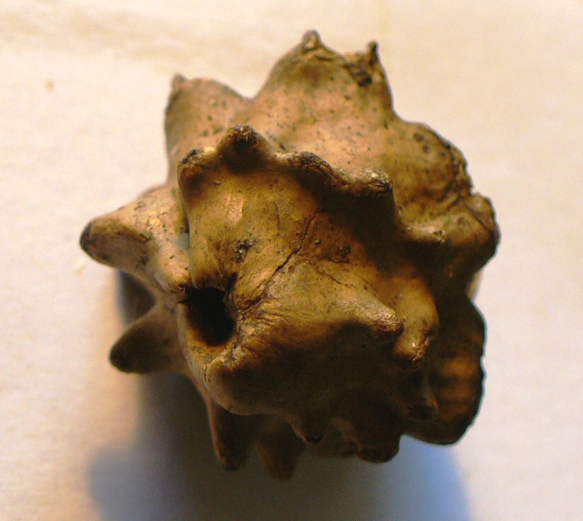
The familiar knobbly form of a Knopper gall (green when fresh, woody when older)
The Knopper grows as a gall on acorns forming a series of protrusions as seen above with an exit hole at the top. Cutting it open however starts to reveal a tiny world of finer structure.

One bisected Knopper...
This shows a thick woody outer wall with an approximately conical chamber inside with thin vertical partial walls or flanges, and the exit hole at the top. Now, looking closer still...

Zooming in on the chamber and one of the protrusions.
Above you can see some other structures - a green-grey mass near the centre and a couple of small yellowish ovals near it (more on these below). Top right is one of the projections and the section shows a pitted texture with a smooth line running through it. I have no idea what this line is - does it provide strength like a laminate or reinforcing bar? Zooming in again shows the pitted structure to be closely packed cylindrical cells.

Cells within the wall of the Knopper gall
Egg case or larval skin?
Even this view has a linear structure running through it - so far I've been unable to find out more, or perceive any deeper structure, but I will keep looking, and welcome any comments on this. Now, the tiny yellowish structures - I think they are either egg cases or shed larval skins (exuviae), but I could see much structure even under higher magnification so have left that for the moment, and a couple of darker structures are what I believe to be frass (insect faeces). The green-grey tufts are a little more interesting though.

Close up of tufts inside the gall
A few possibilities sprung to mind - a lichen, or a microfungus - maybe some sort of mould growing on invertebrate detritus. A quick bit of work with a hooked pin and a microscope slide and the following appeared from the same material:

Filamentous green or blue-green alga

Numerous fungal spores and tiny hairlike structures
From these pictures and the general look of the tufts, I was initially pretty sure this was a lichen - a symbiotic arrangement of fungus (metabolising dead organic matter) and the 'photobiont', a photosynthesising alga or cyanobacterium associated with in. I was thinking of trying to ID the 'photobiont', but a handy comment (see below) suggests that it is a mould along with a separate alga after all (the title stays though!). I've developed quite a few questions from this one common gall, but that's one that may have been answered. Anyhow, that would have been as far as I could get for now, until I noticed a tiny flicker of movement and found this...

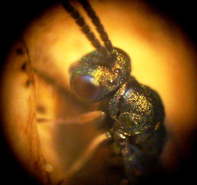
A tiny metallic green wasp

And closer in...
Yes, it is a tiny (2mm tops) chalcid - but not the gall-causer which is a more rotund shape and brownish in colour. This is one of its larval parasites and belongs to the family Pteromalidae. It looks very similar to some other families such as Ormyridae and Torymidae but is distinguished by the narrow pronotal collar, long stigmal vein on the wing and some features of its sculpturing. The genus is uncertain (it really needs a well prepared specimen under high magnification), but is likely to be Cecidostiba sp. Once I've been shopping for the 'Oak Galls of Britain' book (from BPGS's publications page), I may know more.

So, that's where I've got to so far - some interesting observations (I think so, anyway) and plenty of unanswered questions, so as ever, comments and suggestions are welcome. I suspect I'll learn a lot in about a month when the New Naturalist 'Plant Galls' book comes out (already on order...) and will post updates if any of my questions are answered. Until then, enjoy the following gratuitous pteromalid close-ups - a truly lovely little parasite!

Head showing crooked antennae, red-brown eyes with ommatidia (facets) and red-brown ocelli.

Dorsal sculpturing

Iridescent wings and venation

A ventral view, also showing the long stigmal vein


A slightly broader dorsal view.

Wednesday, 9 March 2011

Entomology of Star Wars. Episode III: Bears in Space

Right, here it is - the third and final instalment of 'The Entomology of Star Wars' (unlike George Lucas I will not be tempted to produce a shiny but unsatisfying second trilogy). Following on from Episode II (exogorths - giant asteroid-dwelling worms), this time I decided to focus on the 'mynocks', the parasitic space-dwelling organisms that attach themselves to spacecraft to feed off their energy (they can feed on electrical, electromagnetic and stellar energy, and so may attach themselves to structures such as power cables or 'ion ports'), absorbing it through the suctorial mouth located between their eye stalks. They can also feed on the material of the hull, causing damage or even destruction; in turn they are eaten by exogorths and may survive for some time, flying around inside the worm's gut until finally digested.

A mynock attached to the Millennium Falcon, showing its leech-like sucker. Nice.
A little light reading ensures me that mynocks are silicon-based and fatally allergic to helium (they swell and die); also they lack any major organs and so reproduce by binary fission once they have fed sufficiently. Apparently, there have been other species (or subspecies), including one that had a normal mouth (rather than a sucker), and another that gave birth to live young. They do however have bat-like wings which they can use when in an atmosphere. So, what parallels can be drawn between the mynock and real-world organisms?

Firstly, as I've mentioned before, I'm not going to look at ideas surrounding non carbon-based life - there are plenty of bloggers looking at this already. Similarly, although mynocks can feed on ship hulls, I covered chemosynthesis and the mineral metabolism in Episode II. So, let's start with the most obvious aspect of mynock life (to me anyway) - their ability to survive in the vacuum of space. Although there are no known vacuum-dwelling real-world organisms, there is one experimental example which a lot of regular science readers may well be aware of - the tardigrade. For those who don't already know about this, here goes:

Tardigrades (Phylum Tardigrada) are microscopic to just-visible (adults range from 0.1 to 1.5mm) aquatic segmented animals with eight legs, sometimes known as 'water bears' or 'moss piglets'. They are well known for their ability to survive exceptionally hostile conditions, often more so than any other animal. Tardigrades are able to survive in extreme environments that would kill almost any other animal. Some have survived temperatures down to near absolute zero, up to approx 150°C and about a thousand times as much radiation as other animals, not to mention almost a decade without water. How do they manage this?

Tardigrade - a (tiny) bear in space!


Well, along with some other groups of organisms which can survive essentially complete dehydration, tardigrades accumulate large amounts of disaccharides ('double sugars' i.e. those with two molecular rings), especially sucrose and trehalose (Crowe et al. 1998). It appears that these sugars stabilise membranes and proteins when dehydrated, probably by hydrogen-bonding to polar residues in the dry macromolecular assemblages. This maintains dry proteins and membranes in a state similar to that seen when wet. It has been suggested that, as sucrose and trehalose form a glassy state when dry, glass formation (vitrification) is sufficient to stabilise dry biomaterials - however, Crowe et al. (1998) showed that both direct stabilisation and vitrification are required and that trehalose has properties linked to its crystal structure than may aid the stability and longevity of dehydration-tolerant organisms that contain it

Meanwhile, Jönsson et al. (2005) exposed tardigrades to high levels of gamma radiation. Without going into the results in detail, tardigrades do eventually die and/or become sterile, but the study concluded that their radiation tolerance may be due to currently unknown, but efficient, DNA repair mechanisms (unlike the  biochemical protection seen for their dessication tolerance). Then, in September 2007, tardigrades were taken into low Earth orbit on the FOTON-M3 mission and exposed to either the vacuum of space or both vacuum and cosmic/solar radiation. Despite these hostile conditions, where vacuum causes severe dehydration, while cosmic/solar radiation (with an unfiltered UV component) would be expected to cause considerable genetic damage. However, upon their return to Earth, many had survived and even laid eggs that hatched normally - they are the first animals to survive such exposure (Jönsson et al. 2008).So, they can protect their cells membranes and either protect and/or repair their DNA well enough to not only survive in space, but reproduce afterwards - our first parallel with mynocks, up to a point at least, but are there any others?

Well, I'm fairly sure there are no direct energy feeders (discounting photosynthesis of course!), unless you include the 'mystic woo' of psychic healing etc. Which I don't. However, the binary fission of a large organism is interesting - generally this is associated with small organisms such as Protozoa and Algae. However, it is also seen in some Myxomycetes ('slime moulds', but not Fungi). These are often soil micro-organisms, but in some species a single-celled, but multinucleate, 'plasmodium' is formed which is the final feeding stage, engulfing many kinds of small food items. Although essentially a single cell, these can be large - one example of Brefeldia maxima in North Wales can cover whole stumps, be a centimetre thick over a square metre and weigh up to around 20kg - possibly the largest cell known (Ing 1999), and its certainly feasible that such a structure could divide. So, we have our 2nd real-world parallel.

So, lastly on to the helium allergy... well, helium poisoning certainly can occur (e.g. if overused for 'squeaky voice' purposes), but although there's a general feeling of 'you can be allergic to anything', I've not been able to find anything about genuine helium allergies. It is pretty unreactive, and I suspect at least some of the anecdotal reports I found are actually allergies to the material the helium was held in (balloon rubber, latex glove powder etc). S, although a major anaphylactic event might parallel the effect of helium on mynocks, this remains an unproven 'maybe', and as Meat Loaf said, 'two out of three ain't bad'.

With that, I shall end my 'Entomology of Star Wars' trilogy, but fear not fellow invertebrate-SF nerds - I am starting to form ideas about B-movies and Iain M. Banks novels...

References

Crowe, J.H., Carpenter, J.F. & Crowe, L.M. (1998). The role of vitrification in anhydrobiosis. Annual Review of Physiology 60: 73-103.

Ing, B. (1999). The Myxomycetes of Britain and Ireland. Richmond, Slough.

Jönsson, K.I., Harms-Ringdahl, M. & Torudd, J. (2005). Radiation tolerance in the eutardigrade Richtersius coronifer. International Journal of Radiation Biology 81(9): 649-56.

Jönsson, K.I., Rabbow, E., Schill, R.O., Harms-Ringdahl, M. & Rettberg, P. (2008). Tardigrades survive exposure to space in low Earth orbit. Current Biology 18(17): R729-R731.