Welcome

Welcome to my blog

This is where I post various musings about wildlife and ecology, observations of interesting species (often invertebrates)
and bits of research that grab my attention. As well as blogging, I undertake professional ecological & wildlife surveys
covering invertebrates, plants, birds, reptiles, amphibians and some mammals, plus habitat assessment and management
advice
. I don't work on planning applications/for developers. The pages on the right will tell you more about my work,
main interests and key projects, and you can follow my academic work here.

Wednesday, 25 May 2011

Galls of unknown origin

Those of you familiar with my blog will have noticed a minor fascination with galls lately, in part due to the publication of Redfern (2011). Following a couple of posts about the common Knopper Gall Andricus quercuscalicis, I felt it was time to tackle something distinctly less well known, to be precise, an undescribed species of the gall midge genus Dasineura (Diptera: Cecidomyiidae).

Dasineura ulmaria is a common galler of meadowsweet Filipendula ulmaria leaves, forming reddish-pink pouches with a hairy opening on the underside. However, in recent years (and indeed before, though less commonly), similar galls have been found on dropwort Filipendula vulgaris but with projections and openings on the upper sides of the leaves (see Harris 2010 for details of material seen between 2002 & 2009). As described in Redfern (2011, pp. 147-150), the newly hatched larva of D. ulmaria settles on a young leaf, and as it feeds, nutritive tissue develops beneath and around it, with cells growing and dividing to envelop it. This is typical of much insect-mediated gall formation where larval feeding induces gall production, although the precise mechanisms are porrly understood.With D. ulmaria eggs being laid on the underside of the leaf, the gall also has its opening here, unlike the galls on F. vulgaris as noted above.

Dasineura galls on F. vulgaris
These galls are single-chambered ('unilocular' in gall-speak) and as shown in the photo above, there may be more than one per leaf, and many per plant.

Dasineura gall on a single leaf of F. vulgaris. Note the hole towards the left which may be generalised plant-feeding damage or could be a potential predator of the enclosed larva.
Upper side of a F. vulgaris leaf showing two gall projections and openings.

Close-up of the gall openings showing fringing hairs and elongated cells of the conical projections.
Further investigation of the gall surface shows enlargement of plant cells, especially along the leaf ribs.

Enlarged cells on the outside of the gall, highlighted by reddening/darkening along ribs and veins.
Moving on from surface anatomy of the gall, its dissection reveals the single larva that lives inside.

The tiny (approx. 1mm long) larva in situ showing its yellow colour and segmentation. Also note the somewhat spongy texture of the nutritive cells lining the inside of the gall.

Close-up of the larva (width of photo approx. 0.5mm) showing the small head and tiny (paired) antennae. Also note the segmentation highlighted by dorsal and ventral sclerites, plus a hint of internal structure and gut contents.
In Dasineura ulmaria (and many other galling species), the young larva induces the production of large nutritive cells by the plant which line the gall chamber and provide it with food, especially during later stages/instars. As the larva grows, nutritive cells are consumed and nutrients continue to be supplied from vascular strands and stored starch. As the larva empties successive layers of nutritive cells, replacement nutrients have to pass across several dead layers. In D. ulmaria, nutrients also have to diffuse from vascular strands to the larva via the sclerenchyma - cells which have become somewhat woody but remain alive and connected - and it is possible that this is also the case with the galls on F. vulgaris. The larva pictured above is of the general form expected of Dasineura and although the papillae and structures known as the sternal 'spatula' (underneath the head end) which can be diagnostic (e.g. Smith 1989) were not visible in this young specimen, it is hoped that more mature larvae will be collected during a field visit in June to the same chalk grassland site that produced the specimen photographed here.

As noted by Harris (2010), this gall has been described previously - as D. ulmaria by some authors and D. harrisoni by others, although it may of course turn out to be genuinely undescribed. Therefore, until further work can be undertaken (e.g. on the host range of D. ulmaria and the identity of D. harrisoni which is unclear), this gall is to be treated as distinct but undescribed i.e. Dasineura (undescribed sp. A on F. vulgaris) - evidence that even in over-populated (and entomologist-laden) southern England, new species await discovery!

As ever, watch this space - if there are further developments I hope to be able to post them here.

References

Harris, K. (2010). Notes on gall midge galls recorded on Filipendula ulmaria and F. vulgaris in the United Kingdom. Cecidology 25(1): 6-10.
Redfern, M. (2011). Plant Galls. Collins, London.
Smith, K.G.V. (1989). An introduction to the immature stages of British flies. RES Handbooks for the Identification of British Insects 10(14):1-280.

Sunday, 22 May 2011

What's in a gall? Part 2: home-makers

Regular readers may have seen last month's musings on the contents of a knopper gall of Andricus quercuscalicis. The focus was on the structure of the gall and the presence of the small parasitic wasp Cecidostiba fungosa - this time, a relatively brief look at the gall-causer itself.

A quick pictorial reminder of C. fungosa - a little parasitic green jewel.
I kept the gall to see if any other species emerged, and after a couple more weeks, two individuals of the galling species itself, A. quercuscalicis, emerged - adding to the seven C. fungosa already seen.

A. quercuscalicis
A. quercuscalcicis isn't the metallic green of its parasite - more a dark brown, but interesting nonetheless. The photo above shows the overall form with paler yellow-brown legs and antennae - the latter clearly showing their simple filamentous shape and curvature. The abdomen is clearly rounded and shiny and even in this picture some hints of sculpturing and bristles can be seen.

A. quercuscalicis - close-up of the front half.
Zooming in, the thorax and head clearly bear small pale bristles as well as sculpturing in the form of pits and wrinkles, and the tiny bulbuos base of the forewing is also visible. Just behind the wings (the bottom centre of the photo), there is another clear patch of pale bristles. As ever, such seemingly unassuming invertebrates provide a wealth of visual detail.

However, already knowing the identity of this species as it emerged from a gall in a sealed container, I do not wish to dwell on the finer points of diagnostic morphology (not this time anyway...); instead I'd like to look at the biology of the Knopper Gall. Knoppers are asexual galls and (despite only arriving here in the 1950s) are common in Britain, mainly on pedunculate oak Quercus robur and rarely on sessile oak Q. petraea. Fresh knoppers are found on acorns in summer and autumn, starting green and sticky but becoming brown and woody (like the one illustrated below). The thick wall contains an air space with an ovoid inner gall inside.

An old, woody knopper gall, around 2cm across
The sexual galls of this specis are very different and are found in spring on the male catkins of Turkey oak Q. cerris. Unlike the knoppers, these are tiny (1-2.5mm long) and thin-walled; each one replaces a small number of anthers in the catkin, and a catkin may bear up to around 20 such galls. The galls prolong the life of the catkin slightly as it usually lasts around 12 days while the gall requires 14 days to develop (other gallers may extent catkin life considerably more, by up to three months).

In May the sexual males emerge and wait for females to chew their way out - the males fight to secure the best mating locations. Mated females then fly to Q. robur trees and lay eggs (in late May) into newly pollinated female flowers; each egg is positioned precisely between the developing acorn and cup. In September, the galls drop to the ground (before ungalled acorns) and overwinter, along with the larvae inside, within the leaf-litter. Most (in Britain, 70-80%) of asexual females emerge the following February or March, the remainder staying in the knopper for up to three years and joining a different cohort. In either case, they then fly to Q. cerris trees to lay eggs in male flower buds and begin the life cycle once more.


To finish (for now), I have retained the knoppers and will be interested to see if anything else emerges although it seems unlikely unless further adults remain. So, there may be a third instalment covering internal knopper structure and possibly more inhabitants. I would also like to recommend the following book which has recently been published - it is excellent and was the source for the life cycle summary given above:

Redfern, M. (2011). Plant Galls. Collins, London. [vol. 117 in the New Naturalists series]

Monday, 16 May 2011

Beetle Carnival! An Inordinate Fondness #16

It's been a quiet week or two for me on the blogging front - too much of the paid stuff to do. Still, I knew that for once I had a post booked in advance - issue #16 of the beetle-focused blog carnival 'An Inordinate Fondness'. This is the first time I've hosted a blog carnival (my own blog's not even a year old yet), so where to start?

Well, at the (conceptual) beginning with an excellent post about being drawn into beetling in the Ozarks, the acquisition and building of equipment, and occasional bouts of wifely disapproval - here's Basic Beetling by the Ozarkian. This has a few larger beetles illustrated (though we all know how many are tiiiiny) and mentions the assumption that they'd all be large - like The Perching Dung Beetles of Wongabel by BunyipCo. Apparently, in the Neotropics, dung beetles arrange themselves by size on perches, something that - if you wish to observe it - means sitting out in the rainforest at night; sounds good to me!

Anthocomus rufus (nope not featured anywhere else here!)

Sticking with the dungy theme, plus locations that seem exotic from my location in suburban southern England, the Natural History Museum (to me, the 'Mother Ship') would like to tell us all about the 2nd part of their expedition to Tanzania, this time to the Udzungwa Mountains where it appears they had to Exit, pursued by... a buffalo. Having done plenty of East African fieldwork in my time, I know the feeling... Still, despite the tetchy vertebrates, they found some fine dung beetle specimens using dung-baited traps, and more - including a mystery cerambycid - what more could you want? Well, what about the charismatic scarabaeid Paracotalpa ursina from Sam Wells Bug Page. These were recorded in an ordinary city park in Fresno, showing that witnessing their weighy grass-stem-toppling antics doesn't require a well-funded research trip to a remote 'corner' of the globe.

Hmmm... how about a paper on 'voyeurism in the Coccinellidae'?


Next, and switching families, here's some excellent tiger beetle photography from beetle-friendly ant specialist Alex Wild at Myrmecos (one of my personal favourite invertebrate blogs)? Marvel at the metallic green-ness, fear the mighty mandibles, but you can't avoid the gaze of Alex's Six-spotted Tiger. Also looking at cicindelids, AIF's very own Ted MacRae takes a look at some recently published work on the Rediscovery of Cicindela scabrosa floridana; as with any such rediscovery of something thought to be extinct, there is optimism about it being extant, tinged with concern about its long-term prospects. More research, and hopefully effective habitat management, will tell... In the meantime, don't forget to check out Ted's identification challenges!

Off to a couple of photography sites now, starting with something a little smaller than a dung or tiger beetle - I just love Giraffe Weevils so thanks to Kurt at Up Close With Nature as they don't exist in my part of the world. I was similarly pleased by the Headlight Beetle at Nature Closeups; a Costa Rican click beetle (elaterid) with bioluminescent spots on the pronotum, and something I had never heard of even though there are apparently numerous such species spread across several genera.

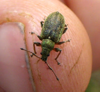
Not a monkey on my back - a weevil on my finger...

Getting back to a bit of 'diagnostic morphology' (one of my favourite aspects of entomology), and finally introducing some carabids which I've so far neglected in this post, Dave Ingram at Island Nature has been searching Vancouver Island for beetles and tracked down a mystery carabid - why not take a look and see what the Backyard Beetle turned out to be. He's also been looking at introduced European species which I am familiar with such as this Gorgeous Ground Beetle.

And finally [insert fanfare here], we return to the Natural History Museum's Tanzanian expedition to try to find out the identity of their mystery cerambycid at And the beetle you have been waiting for... Will it be identified, will it remain a mystery? Head over to find out, and maybe check out their latest video offering too.

One of my specialist group, the Chrysomelidae - leap, tiny flea beetle!
And so we finish - I decided to illustrate this post with a few fairly random beetle photos of my own - better photographers than me are featured in the carnival links above, but please do have a browse around my site. It's not all beetles, and some posts are even about things with bones, though invertebrates do play a major part. I hope you enjoyed this carnival; next month's AIF#17 will be at Biodiversity in Focus so don't be shy - submit a post or two, and maybe even host (it's quite easy) - it's what makes the carnival go round!

Wednesday, 4 May 2011

Stings 'n' things - wasps in close-up

After some recent bee-related posts, I thought I'd stick with the Hymenoptera theme for a while and take an uncommon look at a common species, the Common Wasp Vespula vulgaris. First of all, it's important to know how to identify this species - after all it is quite similar to other social wasps of the family Vespidae.

Wasp-face showing the 'anchor' mark between the eyes.

The thorax - note the four yellow dots forming a square between the wing-bases.


So, looking at the wasp-face above, the 'anchor' mark is a good clue - this is found in V. vulgaris and the closely related Red Wasp V. rufa. However, the latter only has two yelow dots on the thorax, so this is indeed the Common Wasp.

Identification aside, I wanted to take a closer look at an often overlooked (and sometimes unpopular as it is defensive of its nest and does sting, and may number into the thousands in a nest) invertebrate. The face clearly shows tufts of hairs, plus punctures on the mandibles, and the antennae have a velvety look to them with the segments only indistinctly separated. Another feature used to differentiate vespid species is the 'malar space' - the gaps between the bottom of the eye and the top of the mandible. In both the Common and Red wasps, this is short, but in some species such as those in the genus Dolichovespula, it is noticeably longer. However, taking a tour of our wasp means starting with larger structures, so here's a side view of the head and thorax, plus one of the abdomen.
Wasp head and thorax in side view
Wasp abdomen in side view
Here we can see tufts of hairs on various structures - the head including behind the eye, leg segments, and on much of the thorax and abdomen. The eye is indented around the antennal base, and the antennal segmentation is more clearly visible. We can also see the overlap of abdominal segments. So, taking a closer look still and moving forward from the rear, what can be seen?
Tip of wasp abdomen
Here we see the tip of the abdomen through which the sting can protrude - the very tip has a couple of tiny protrusions at the lower edge which may help guide the sting, but no other specialised structures are visible.
Close-up of abdominal segment
Looking at the abdomen more closely, it is clear that, as in many insects, the chitinous surface is covered in tiny pits and between these is even finer sculpturing. Such pits or 'punctures' are well-known in beetles - for many species, the lines (or random arrangement) of punctures on the wing-cases (elytra) are used in identification. However, although rarely looked at in wasps, the same structure is present. In some insects, some pits bear tiny bristles, but I have to wonder if they perform a weight-reducing function, somewhat like a honeycomb structure on the outside.
Underside of the mid-section
Underside of the waist in close-up
The mid-section of course has the familiar 'wasp-waist' (legs bases can also be seen just in front of it), but zooming in, the underside of the waist has a shallow groove bearing yet another tiny tuft of hairs. What function this has I can only speculate, but maybe it accomodates the curve of the abdomen when this is brought forward with the hairs providing a sensory function. Coarser, longer hairs are visible on either side.
Tarsal segments

The 'foot'

Close-up of the wing
The general structure of the leg can be seen in the side view nearer the top of the page, but here the tarsi and the foot itself are seen more closely. The tarsal segments bear tiny bristles and stouter spurs and the articulation between two tarsal segments is visible. The presence of spurs on hymenopteran legs is not unusual and may form apparatus for cleaning the antennae. The foot itself has the typical arrangement of a pair of curved claws on either side of a central pad, again with tiny hairs and a few larger bristles - I imagine the functions are split between sensory use and improved grip. The wings are folded longitudinally at rest, and here numerous tiny hooks are visible on the surface, these may help the wings stay together Velcro-style, and there may also be a function in aiding airflow during flight.
The top of the head showing ocelli

Close-up of the mandibles

Close-up of the eye and antenna
Finally we return to where we started - at the head. The top of the head bears small simple eyes (ocelli) while the compound eyes provide more detailed vision via the numerous facets (ommatidia) shown above. The velvety texture of the antennae is revealed to be due to the structure of the surface - numerous tiny holes and protrusions involved in the functioning of this highly sensitive organs able to detect a range of important chemical cues relating to reproduction, feeding and of course the social behaviour of this nest-building species. The mandibles are serrated and well able to chew both food and the wood required to make nest-paper for its predominantly underground nests which are usually in sandy soil although it does sometimes use roof-spaces (Allen 2009). Even the mandible shows tiny hairs and although smooth and hardened, some microstructure as seen on the abdomen (along the darkened cutting edge), plus some tiny pimples. So, we have a common species, but one which provides an excellent example of insect structure and its relationship with function - I guess this is why there are textbooks with titles such as 'Biology - Form & Function'. Also, it's worth remembering that in the last 20 years or so, even this common species appears to have declined in Britain, and in some years can be surprisingly scarce (Baldock 2010). Hopefully the beauty of its structure can help develop a greater appreciation of even our less popular insects...

References

Allen, G. (2009). Bees, Wasps and Ants of Kent. Kent Field Club.
Baldock, D.W. (2010). Wasps of Surrey. Surrey Wildlife Trust, Woking.