Welcome

Welcome to my blog

This is where I post various musings about wildlife and ecology, observations of interesting species (often invertebrates)
and bits of research that grab my attention. As well as blogging, I undertake professional ecological & wildlife surveys
covering invertebrates, plants, birds, reptiles, amphibians and some mammals, plus habitat assessment and management
advice
. I don't work on planning applications/for developers. The pages on the right will tell you more about my work,
main interests and key projects, and you can follow my academic work here.

Monday 27 June 2011

Eight legs good

Several of my post in recent months have been on the topic of 'diagnostic morphology' i.e. taking a close look at invertebrates (some common, some not) to see why they are what they are.  I've looked at beetles, galls, wasps and others, but have so far written little about spiders. So, when I found a colourful little spider drowned in the garden yesterday, it seemed only fitting that its demise should be of some value - though it wrinkled once dry, the key features remain visible.

Dorsal view of the dried specimen of Enoplognatha ovata
From the first photo, the abdomen shows the distinctive green-yellow colour with a central broad red stripe, although this is variable - the stripe is often missing with just some small dark dots instead. The carapace has a thin central dark stripe and the whole spider (excluding legs and other appendages) is about 4mm long. These features alone allow identification as E. ovata, but there are other features such as the large divergent (spreading) chelicerae (jaws) bearing long fangs. These are seen in males of the genus Enoplognatha, along with the 'boxing glove' palps used in reproduction. Let's look in a little more detail.

Ventral view - the palps and fangs can be seen clearly, plus the leg attachments underneath the thorax - which also shows the thin central dark line visible on the dorsal surface.

Dorsal view of the abdomen detached from the thorax. The point of attachment is bottom left and is surrounded by small parallel stripes - probably muscle attachments. The rough ovals are the leg attachments visible through the abdomen.

Side view of the abdomen - this shows the mosaic colour pattern which appears uniform from further away. Also, what appears to be a smooth abdomen is shown to be bristly under higher magnification.
E. ovata is a member of the family Theridiidae, also known as the 'comb-footed spiders' and it is worth looking at some features that show its place in this group which tends to build random, tangled three-dimensional webs.

The tip of a leg showing not only the bristles, but also the 'comb-foot' that gives the family its common name. These are used to comb out silk from the spinnerets during web-building - this silk is not sticky, but entangles prey.

The arrangement of eyes (the front is to the right). Relative to the small size of the spider, these are fairly large and protruding. Apart from the front middle eyes (which are always dark), these tend to look pearly when the spider is alive. Note the fusion of the side pairs of eyes. Also, there are a few tiny bristles present on the head. This arrangement of eyes with two short rows, the side pairs fused, is typical of the Theridiidae.

Close-up of the fused left-hand pair showing them protruding, and the wrinkled structure where they join. Below this some faint dark lines are just visible internally which may be nerve fibres running from the eyes.
Lastly I'd like to look at the appendages attached to the underside of the head - the chelicerae and palps.

In the left, the male's oval palp. On the right the large fang of one of the chelicerae. In other genera within the Theridiidae, the chelicerae are no more than medium-sized. Here the central channel is just visible running through the centre of the fang - this family of spiders has a fast-acting venom which, though they can't bite humans, quickly disables its invertebrate prey.

The male palp.The structure varies by species and receives spermatozoa from the testes. It is then used to transfer sperm to the female. In adult male spiders, the palps are the most important feature needed for identification to species (where this is not otherwise straightforward). In females, it is the epigyne, the area around the genital opening which receives sperm from the palps. Here note the bulbous end segment of the palp forming a 'cymbium' within which the palpal organs (the reddish protrusions etc) are located.
As in previous posts of this type, I hope this helps clarify the importance and function of fine structure - sometimes it is required for identification, but in any case is interesting as it illustrates how 'form and function' are inextricably linked.

References

Jones, D. (1989). A Guide to Spiders of Britain and Northern Europe. Hamlyn, London.
Roberts, M.J. (1995). Spiders of Britain and Northern Europe. HarperCollins, London.

Wednesday 22 June 2011

Feathers, flowers and feelers - more on wildlife-friendly gardening

Back in late March, I posted about bee-friendly gardening and the range of species found in our little terraced plot in southern England. Three months on, the early summer has turned into autumn, back to spring, and may again become summer soon, so I thought I'd provide another informal update about what's been happening in the garden. Also, Blogger's not playing nicely today, so apologies for the peculiar formatting 'choices' below!


As I sit here, I can see our contribution to the (hopeful) recovery of the UK's beleagured House Sparrow (Passer domesticus) population flitting about in the garden near the seed feeder - including fledged young still fluttering for food near their parents. There is no evidence of sparrows nesting in the garden, but just outside the front of our house, they can be seen being particularly resourceful...


Female sparrow sitting outside a nest entrance - inside a telephone-line junction box. The male has also been guarding and there has been much activity with food and nest materials being brought. This box was also used last year, but the box on the other (south-facing) side of the pole is much less popular - maybe due to the temperature difference?
Of course, sparrows are not the only birds using the garden - the bath has been very popular with bickering, and occasionally even English-style queueing.

Blackbird (Turdus merula) drinking from the bird-bath


Wood Pigeon (Columba palumbus) hogging the bird-bath - the passerines can be heard shouting nearby...
Now, our garden isn't huge - it's a long thin Edwardian terrace; there's hard-standing at either end (sheds, patio etc) but in the middle it's split into flower & veg beds, plus a wild 'meadow' area. It may not be huge but it does have diversity of structure, plus the planting is either native or known to be wildlife-friendly (especially for pollinators). Also, even the hard surfaces have their purpose - plants are allowed to grow over the sheds, and on the patio, some isolated slabs have been removed to allow low-growing, mat-forming species to be planted.


Meadow Clary (Salvia pratensis) - extremely rare in Britain, we now have one plant in our tiny wild meadow! Hoping to collect seed...
An ant's-eye view of the burnet-like Acaena planted where a patio slab has been removed.
There's more I could add about birds and plants, but as you may know, my favourite group is the invertebrates. Now, I do understand that many gardeners may be suspicious about non-pollinating insects and consider many to be pests. However, apart from a very few (we do apply nematodes as a biological slug control), we tend to leave the plants and beasties to fight it out among themselves and have had no major problems so far (the pigeons are more troublesome). It also provides opportunities to watch some interesting inter-species interactions.


Partly added as a pretty picture, here is a spider (Metellina sp. I think) building a web inside a foxglove (Digitalis purpurea) flower, hoping to catch those flighty nectar-feeders.
One of our many many garden ants (Lasius sp.) tending, maybe 'milking' honeydew from, A Brown Peach Scale insect (Parthenolecanium corni).

The diversity of arachnid forms writ large - a Trombidium mite and a Pardosa wolf-spider
While on the subject of spiders, our garden is full of Garden Orb-weavers (Araneus diadematus) which implies there's plenty of food - unsurprising given the array of tiny things that crawl and fly here. However, it was only recently that I saw the stage in their development just prior to scattering (somewhat literally) to the four winds.

Shhh... the spiderlings are sleeping...
What was that? They've started to stir...
Aaaagh, run for it!
Some bungee from the bottom of the nursery web, others make a run for it - the one top right is winning...
Oh, it wasn't anything dangerous - they start returning to the web to reform the ball, some climbing back up bungee-ropes, others crawling back along various strands. If it had been a predator or other threat, the chances are some would have escaped as they scatter in all directions.

And so, there is space for a wide range of species even in our fairly small garden - it just needs some thoughtful planting, a few other features (feeders, bird-baths, a pond is in the construction stage), diversity of structure, and a willingness to leave things alone as much as possible. This is an important little bit of nature conservation most of us can do to some extent, and if you enjoy recording species, gardens can be important sources of biological records (in the UK there are popular garden-based bird and, to a lesser extent mammal, recording schemes designed for non-specialist volunteers). Anyhow, enough from me today - more soon, and eventually I imagine I will eventually compile a garden species list...

Tuesday 21 June 2011

What's in a gall? Part 3: Secrets of the bedeguar

About a week ago, I was on top of St. Catherine's Hill nature reserve near Winchester (Hampshire, England), leading a wildlife walk entitled 'Galls and other wildlife' as the heavens opened and the rain came down... and down... However, although few invertebrates were visible, there was one aspect of entomology that was visible whatever the weather - galls. Last month, I wrote about an undescribed gall species which can be found on this chalk grassland reserve, and before that I investigated the complex inner workings and inhabitants of a common gall species, the Knopper. So, when I saw a cluster of old woody bedeguar galls (Diplolepis rosae) on a sweet-briar (Rosa rubiginosa), I had to collect one and bring it back for closer examination.

A live gall of D. rosae


Bedeguar galls (also known as Robin's Pincushion in Britain) are the galls of the cynipid wasp D. rosae and are quite familar due to their large (a few cm across) spiky shape as shown above. Bedeguar is a Persian word relating to thistles (either their spininess or being wind-blown like thistledown), and there is evidence that ancient writers such as Pliny were familiar with these structures.

Most (well over 90%) of D. rosae wasps are female and it may well be that males are redundant and disappearing. As described in, for example, Csoka et al. (1998), this appears to be due to infection by the bacterium Wolbachia which causes reproduction to occur via thelytokous parthenogenesis (i.e. production of females from unfertilised eggs). Wolbachia has various effects on its different hosts (other wasps, woodlice, gnats, fruit flies) and comes in different strains, but it can interefere with meiosis (meaning females could not produce haploid eggs), cause sexual incompatibility, prevent production of males, or even feminise males (this happens in woodlouse hosts even though they have males genes). In any case, females emerge during May and June and lay eggs in leaf buds which are beginning to swell - whatever the precise criteria for bud selection, females can investigate a bud for up to an hour before deciding on its suitability. Inserting the ovipositir under a bud scale, eggs are laid between the developing leaflets inside  without damaging the plant tissues, and 30+ eggs can be laid, each in an individual cell.

As is usual in cynipid gallers, the leaflet cells around and below the egg immediately become highly active, enlarging and producing RNA, proteins and other substances, and a small pad is formed after about two days. Cell walls break down forming a cavity in the pad, and around a week after the egg was laid, it hatches and the larva enters the cavity. As it begins to feed, nutritive cells develop near its head - these line the chamber while the outside produces cambium. The epidermis meanwhile grows bulges which develop into the familiar multicellular hairs forming the outside of the gall which is familiar to us, the whole mass containing a number of cells. Also, new vascular tissues grow inwards to supply the nutritive layer and out into the hairs as well as linking with leaf veins. The gall is fully developed by July or August and at this point the larvae feed rapidly, being fully fed by October. They overwinter in the gall, pupate there in late spring, and new adults use their jaws to tunnel out (Redfern 2011).

An old bedeguar gall c. 25mm across showing exit holes in individual cells. The gall is also covered with lichens and moss.

The gall broken open to show vascular strands and the inside of a single cell.
The pictures above, especially the lower one, show the complex structure of the vascular tissues, shown here as spaghetti-like threads having grown through the outer layers before they became woody. As shown by the colonising lichens etc., this is quite an old gall and so no D. rosae or associated parasites/inquilines are present, but the complex structure is of interest, especially given the possibility of non-galling invertebrate colonists. A variety of structures is shown in the following pictures:

Enlarged cells similar to those seen in the Knopper gall (see link at top of article)

Layering of cells in the bedeguar gall.

Spongy texture of woody cells surrounding a gall cell.

Some of the vascular tissues, now woody, linked to various parts of the gall and host plant, including the hairs - elongate plant cells are visible.

A section from around a gall cell showing the same elongate structures and their association with the spongy layer.
Looking inside an individual gall cell, it is clear that there is colonisation by, for example, lichens and fungi, but there is evidence of other biota using these ready-made structures.

The inside of a gall cell showing a white membrane suggesting a cocoon, plus black specks of 'frass' (invertebrate faeces).
Looking behind this membrane, among the green algae and black frass, there appears to be an empty skin (exuvium) - the small linear structures in the centre are probably legs.
Having found an exuvium, I had to wonder what had left it there. I didn't expect to find anything, but then there was a tiny flash of reddish movement as I looked down the microscope.
In the bottom of the open cell, a small, round (and quickly moving) red shape.
After some time, I managed to capture this tiny creature (the inner chamber of the cell is only a few mm across, so this is only about 0.5mm long) and took some pictures. It turns out that unlike many invertebrates, it is unable to walk on glass, so although its limbs were moving, the organism itself stayed still on a slide...
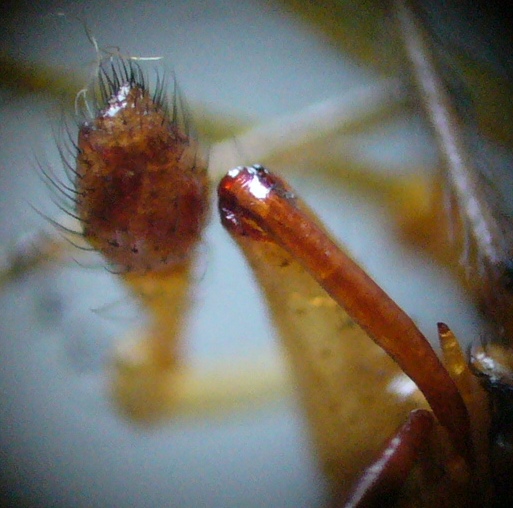
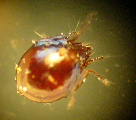
The tiny beast in question - a mite, possibly an oribatid. Note the shiny round carapace, bristly appendages and rostrum with transverse wrinkles.

The dorsal surface of the mite showing the even sculpturing.

The best close-up I could get of the head and front appendages/bristles.
So, as always when I decide to investigate a gall in detail, I have come away finding more than expected - it has induced me to read up on the unusual bacterium-mediated reporductive strategy of D. rosae, scrutinise the fine structure of something that is familiar on a macro scale only, and find an invertebrate that I genuinely can't identify (I await a friendly acarologist - should one appear, I'll post any updates). However, with its shiny single carapace, it does look like an oribatid (moss mite, order Oribatida). These vary in their diet, but different species feed on dead plant matter, fungi, carrion or lichens, while some are predatory. Given the microhabitat here, I suspect fungal and/or lichen feeding, and after a further search of the gall cells I found four of these mites. The gall and mites are now in a tiny vivarium, so I may get to see more behaviour and maybe young. If so, pics and details will of course appear here. Thanks for reading!


References

Csoka, G., Mattson, W.J., Stone, G.N. & Price, P.W. (eds) (1998). The Biology of Gall-Inducing Arthropods. General Technical Report NC-199, Forest Service, North Central Research Station, USDA, St Paul, MN. Contains many useful papers and used as a general reference in the publication below.

Redfern, M. (2011). Plant Galls. Collins, London. The source of much of the gall biology here, and a must for gall-nerds!

Saturday 18 June 2011

Jewels in the reeds

My last post - a photographic offering from a local water-meadow site - included a picture of a reed beetle and a promise of something more in-depth on this group of invertebrates. So, here it is...

First of all, what are reed beetles? They are the subfamily Donaciinae within the Chrysomelidae (leaf beetles), and are medium-sized (5-12 mm), fairly elongate and often metallic beetles found generally on waterside vegetation. In Britian there are 21 species within three genera - Donacia, Macroplea and Plateumaris. However, despite being of a fair size with charismatic colouration, some are highly variable in colour whilst true morphological differences between species can be slight. This, coupled with a lack of accurate illustrated keys, meant that there was considerable confusion regarding identification of this group in Britain prior to the publication of keys by Menzies & Cox (1996), and subsequently (currently undergoing revision) Hubble (2010). Knowledge of the group was also greatly enhanced by the publication of an illustrated and annotated atlas (Cox 2007). A visit to an old Hampshire water-meadow in late May 2001 produced many sightings of several species and it is these that are presented and discussed here.

The River Itchen as it runs through the water-meadows.
Apart from being most picturesque, the photo above shows a small path alongside the River Itchen and it is the fringing vegetation (reeds, irises, sedges, Sparganium etc.) for several hundred metres that was the source of the specimens here. Identification of the Donaciinae relies on a range of features - sometimes colour is helpful, but often closer examination is required of features such as pronotal puncturation and sculpturing, femoral spines, antennal segmemts and sometimes dissected genitalia. Therefore, although I have attempted to make definite identifications to species, these were not collected (it was initially a day of voluntary conservation work rather than bug-nerding), and hence - relying on photographs and field observations, despite being in the middle of 'writing the book' - some identifications are tentative. Still, I hope the material is interesting...

A pair of reed beetles on Iris pseudacorus
The pair of beetles illustrated above gives a good indication of their charismatic nature and shows some key features despite them being tricky to ID in the field - the overall shape, metallic colour and 'bumps' towards the front corners of the pronotum (not present in all species). These are (I think) Donacia semicuprea - note the midline of the pronotum with two unequal-sized dimples (fovea; pl. foveae) joined by a groove, and the elytra (wing cases) which are slightly rounded and widened in the front half. The pronotum and elytra are the same copper-bronze colour, and although it is hard to see in this picture the sides of the elytra have a faint greenish tinge - the head is greyish due to short pale pubescence. There are femoral teeth visible which are usually not present, but this is a variable feature and it is not that unusual to find them.

A green reed beetle on a yellow iris flower

A blue reed beetle running along the stem of an umbellifer

Dorsal view of a blue reed beetle
These three images show a quite different colouration - metallic blue and green - and the middle picture tries to illustrate the 'vaulting' (as opposed to relative flatness) of Plateumaris compared with Donacia in side view. Again, this is trickier than it feels it should be, but I think these are all Plateumaris sericea - the variable colour is well-known, and the top picture shows the strong thorn-like tooth on the hind femur. Also, this species can be separated from the similar P. discolor to quite an extent by habitat - P. sericea is found in neutral to high pH areas (as found at the water-meadows which are on chalk), while P. discolor is found in more acidic conditions.

A metallic-green reed beetle
And so to the third and last species that I will be treating individually here. Donacia simplex is arguably the most common British reed beetle and is characterised by the 'frosted' metallic coppery colour, often with a uniform tinge of green (as here), red or grey, plus at least some orange-red on the legs and antennae (faint in the photo, but clearer in the field). Also, the femora are untoothed and it is generally found on Sparganium bur-reeds (this is on a different plant which was among a Sparganium clump).

So, what of the rest? Well, the following pictures are the 'best of the rest' - generally unidentified, or only identified tentatively, due to my unwillingness on this occasion to collect them - some jewels are best left unmined and I had no urge to add them to an entomological collection. Instead they are here to show the varied nature of these splendid beetles. Enjoy!

A reed beetle on a curled iris petal - unidentified, but the pronotal groove and faint elytral green-gold banding suggests Donacia marginata as the usually clearer marking are variable and may be reduced or absent.


Two images of another charismatic specimen - this time with a reddish tinge on the pronotum and front third or so of the elytra.

Two specimens of what I think are the same species with shiny, almost sparkling, reddish-brown to dark brownish-green elytra.
 
And last, but not least - the Donaciinae - feel the love!

References

  • Cox, M.L. (2007). Atlas of the Seed and Leaf Beetles of Britain and Ireland. Pisces, Newbury.
  • Hubble, D. (2010). Keys to the Adults of Seed and Leaf Beetles of the British Isles (Coleoptera: Bruchidae, Orsodacnidae, Megalopodidae and Chrysomelidae). Test Version. FSC, Preston Montford.
  • Menzies, I.S. & Cox, M.L. (1996). Notes on the natural history, distribution and identification of British reed beetles. British Journal of Entomology and Natural History 9: 137-162.
Further sources

For excellent online coverage of the European Donaciinae, including many photos of prepared specimens, see here.

Thursday 2 June 2011

Water meadow wonders

As summer seems to have returned to southern England, I thought I'd offer something a bit lighter and essentially photographic rather than any in-depth diagnostic morphology or analyses of gall contents (though these will return...). Instead, here's a selection of splendid invertebrates from Ovington water meadows near Winchester - the site is managed for nature conservation and is a centuries-old water meadow system of sluice-controlled channels, grasslands, scrub and mill-stream. Enjoy!

Starting with the flies (Diptera):

The Noon-fly (Mesembrina meridiana) often seen basking - distinctive yellow-gold wing-bases on an otherwise black fly. This one was busily engaged in leg-cleaning behaviour.

A hoverfly of the genus Helophilus feeding from umbellifer flowers.

The stilt-legged fly Calobata petronella perched on a leaf.

 And of course some beetles (Coleoptera):


A splendid Red-headed Cardinal beetle, Pyrochroa serraticornis

An (IMO) even more splendid longhorn beetle, Agapanthia villosoviridescens

Guarding its patch of umbellifer flower, a soldier beetle of the genus Cantharis.

One of the Donacia reed beetles - in my specialist area, the family Chrysomelidae. Look out for a post on Donacia soon, including colour variants from this site.
 Now onto a possibly less familiar group, the scorpion-flies (Mecoptera):

A female of the genus Panorpa - the males have a scorpion-like tail with genital capsule raised at the end.
 And finally, I couldn't look at a wetland ecosystem without at least one specimen of Odonata:

To humans, a lovely Banded Demoiselle Calopteryx splendens; to small invertebrates, fearsome flying jaws!
I hope you enjoyed my photographic interlude - something a little more in-depth coming this way soon...