Welcome

Welcome to my blog

This is where I post various musings about wildlife and ecology, observations of interesting species (often invertebrates)
and bits of research that grab my attention. As well as blogging, I undertake professional ecological & wildlife surveys
covering invertebrates, plants, birds, reptiles, amphibians and some mammals, plus habitat assessment and management
advice
. I don't work on planning applications/for developers. The pages on the right will tell you more about my work,
main interests and key projects, and you can follow my academic work here.
Showing posts with label defence. Show all posts

Thursday, 19 November 2015

Stinky sticks

I keep black beauty stick-insects, Peruphasma schultei which hide, not by looking like vegetation but by being black, velvety (hence non-reflective) and nocturnal. However, although usually calm when handled, they do have active defences which I discovered recently when retrieving a large females that had escaped to climb the curtains. I felt something wet on my hand, and when I looked there was a milky, eye-wateringly acrid liquid. I knew that a lot of stick-insects can spray defensive chemicals but hadn't witnessed this one before. Fortunately a quick Google told me that this had been investigated by Dossey et al. (2006), who discovered a new defensive compound that they named 'peruphasmal' after the insect producing it.

Peruphasma schultei
Peruphasmal is an isomer of dolichodial, a compound in the iridoid group.

structure of dolichodial
Iridoids are found in many plants (usualy in the form of glycosides) and may be active ingredients in those used medicinally. They are also likely to have a defensive function, protecting the plant against herbivores. Dolichodial and its isomers are found in plant essential oils, and as in this case, in the defensive sprays and secretions of some insects, possible being sequestered from food plants, or being produced by the insect's metabolism (they are intermediate compounds in the production of alkaloids for example). My P. schultei are fed on privet (Ligustrum sp.) which is in the family Oleaceae, one of those known to produce iridoids, so sequestration is plausible. The toxin in Ligustrum appears to be the glycoside syringin, known originally from lilacs, but now known to be a white, crystalline, bitter toxin in many plants including privet, hence its alternative name 'ligustrin'. So, this is a likely candidate for the source of the insect's defence. In plants, iridoids are usually bound to glucose, and peruphasmal is also sprayed along with glucose. Whatever the source - there is more research, but not much - it is an effective defence; my reaction was to put the insect down and wash off the secretion - a predator would have been sprayed in the face...


References

Dossey, A.T., Walse, S.S., Rocca, J.R. & Edison, A.S. (2006). Single-insect NMR: a new tool to probe chemical biodiversity. ACS Chemical Biology 1(8): 511-514.

Saturday, 9 August 2014

March of the green vomit-grubs

As you may know, I am a stakeholder in a nearby community farm, here in the sometimes-sunny south of England. We grow many different crops and one of these is a small patch of asparagus. So, I was interested to see some beetle larvae eating the leaves and stems. Fortunately they don't seem to be affecting the asparagus growth noticeably so they've been left where they are (we don't use pesticides but could remove them by hand if they become a problem). Handily, I specialise in chrysomelids (leaf beetles) and recognised them as larvae from this family: the small shiny head capsule, and the body widening towards the rear then coming to a blunt point is typical. I've been looking at writing a guide to juvenile chrysomelids so in this case, knew they were larvae of the asparagus beetle Crioceris asparagi, and the presence of the distinctively colourful adults confirmed it.

Larvae of Crioceris asparagi along an asparagus stem. Adults in the background.
Late instar larva of C. asparagi eating an asparagus stem. This mature larva is close to pupating and shows a typical chrysomelid larval shape (though some differ greatly especially the tortoise beetles in the subfamily Cassidinae).
Larvae such as this are clearly potential prey for insectivorous predators that like juicy grubs. However, while some chrysomelid larvae hide and protect themselves with shields of their own faeces and shed skins, this species has a different approach. When threatened, it arches backwards, raises its head and regurgitates a droplet of partly digested food onto its attacker. This might not do much to a human but could be noxious to a smaller organism, causing the larva to be avoided or giving it time to drop off the plant to safety.

C. asparagi larva with a regugitated droplet on its head.
C. asparagi larva having used its droplet against my threatening finger.
In contrast, a chrysomelid larva (unidentified) with a shield of faeces and shed skins.
Assuming the larvae survive to pupate and emerge from their soil cocoons as adults, their defence changes greatly as they are colourful, warning potential predators that they are (or in the case of this species, are pretending to be) toxic - something known as 'aposematic mimicry'.

C. asparagi adult.
C. asparagi adults creating the next generation.
So, for now I'll enjoy these interesting beetles - hopefully they won't become too numerous...

Wednesday, 23 April 2014

Springtime beetles, legs go spring

Having a garden pond means occasionally finding dead things floating. No, not hedgehogs or other larger fauna - the sides are profiled so that mammals can get out - but sometimes incautious invertebrates drown. Naturally, if they look interesting (and haven't been eaten by pond skaters and other small predators), I fish them out for a closer look. When I saw a flea beetle (in the Chrysomelidae, my specialist group - they are in the tribe Alticini within the subfamily Galerucinae) I identified it easily enough as Longitarsus pellucidus, a common species, though it's always worth checking. It lives on bindweeds (Convolvulaceae) which are growing near the pond so presumably jumped into the water by accident when disturbed by a spider, bird, cat, human or other potential threat. However, it did give me an opportunity to look at why flea beetles are so named.

Longitarsus pellucidus - the swollen himd femora can be seen, as well as the elongate tarsus that gives it its generic name.
The hind femur in flea beetles is swollen to accomodate jumping muscles and the 'metafemoral spring' which is visible through the cuticle as a slightly paler comma-shaped structure taking up much of the inside of the femur.

Hind femur and metafemoral spring of L. pellucidus.
The spring is a long-oval chitinous structure coiled much like a loose fist. Muscles squeeze the spring closed and then a 'catch' is released and the stored energy is released, extending the leg so the beetle can jump.Here you can see a ligament extending from the spring to the tibia so the leg is folded when the spring is compressed. This mechanism differs from that in fleas which have a blob of resilin (a natural rubber) which is compressed instead of a spring. Although fleas may be more familiar for their leaping ability, some alticine beetles are actually able to jump comparatively further, and of course some of these beetles are no bigger than a large flea - smaller beetles jump further relative to their size (Schmitt 2004). Also, the spring may be useful for identification and for grouping species taxonomically by looking at similarities and differences in the form of their springs. However, this is not a mainstream technique, largely because of the difficulty of dissecting out the spring and the lack of readily available published material forming a guide to identification this way, although there is useful information in Furth (1988) along with images and drawings from various species.


References

Furth, D.G. (1988). The jumping apparatus of flea beetles (Alticinae): the metafemoral spring. In: Biology of Chrysomelidae, eds. P. Jolivet, E. Petitpierre & T.H. Hsiao, pp. 285-297. Kluwer, Dordrecht.
Schmitt, M. (2004). Jumping flea beetles: structure and performance (Insecta, Chrysomelidae, Alticinae). In: New developments in the Biology of Chrysomelidae, eds. P. Jolivet, J. Santiago-Blay & M. Schmitt, pp. 161-169. SPB, The Hague.

Sunday, 29 December 2013

Highlights of 2013

December's been a quiet month on the blogging front - a large beetle project is ongoing (status review of the UK Chrysomelidae) and then of course the whole festive-season-thing. However, there was a proper summer this year with an extended period of hot dry weather extending into a mild autumn, and this meant some fine invertebrate (and other) sightings after some truly awful, cool, wet summers. The most spectacular (for me as they were all personal firsts) were probably three Lepidoptera finds between July and September - two butterflies, a monarch (Danaus plexippus) and long-tailed blue (Lampides boeticus) and a moth, the Clifden nonpareil (Catocala fraxini). The monarch is a North American species, and although some have been known to cross the Atlantic, it is more likely that this (and one from a nearby friend's garden) had escaped from a butterfly farm, maybe on the Isle of Wight. Certainly there was a small flurry of records of this species in southern England, aided by the fact that monarchs in the UK often visit gardens to seek their foodplant, milkweed (Asclepias syriaca) which is of course also non-native. The other two are scarce migrants seen in higher-than-usual numbers due to the favourable conditions this year. Being native to NE Europe, the Clifden nonpareil is more often seen on the eastern coasts of Britain, but my sighting was in Hampshire, about 10km inland where one large and unmistakable adult was seen basking on warm brickwork near scrub including its foodplants - aspens and other poplars (Populus spp.). Also a rare migrant, the long-tailed blue can be found on various Fabaceae such as everlasting-peas (Lathyrus spp.) and brooms (Cytisus spp.) - as a Mediterranean resident, it's not often seen in this country. I'll stop there, but if you'd like an affordable and user-friendly guide to European butterflies, one of my favourites is Haahtela et al. (2011). More to come from me in 2014, but until then, here are some pics from 2013...

A flock/mob of jackdaws in spring, as seen from my study window.
Adult female smooth newt in our garden pond.
A leaf beetle larva and its defensive shield of faeces and shed skins.
And finally, just to prove that I do go out and do ecology in the field, here I am taking great created newt eDNA samples at Claylands Nature Reserve, Hampshire.

Reference

Haahtela, T., Saarinen, K., Ojalainen, P. & Aarnio, H. (2011). Butterflies of Britain and Europe: A Photographic Guide. A & C Black, London.

Wednesday, 27 November 2013

The spider with emerald jaws

As well as mystery cocoons, breaking up some old fence panels for firewood dislodged numerous scuttling invertebrates. Plenty of woodlice, barklice and so on, and many small spiders, but also a splendid specimen of Segestria florentina. This is the largest species of the familt Segestriidae in Europe, with females reaching up to 22mm in length (excluding legs and other appendages). The family is distinguished by having 6 eyes arranged in a semi-circle (most spiders have 8) and the first three pairs of legs directed forwards (most have the first two forwards and the other two backwards). Although this species can bite (it's apparently painful, a bit like a bee sting or sharp jab with a pin, but not dangerous to humans), when disturbed, they curl up or flee to find a crevice to hide in - being nocturnal hunters using tunnel-webs with radiating threads. First found in Britain in the 19th century, this is a circum-Mediterranean/continental species that most likely arrived with ships to ports in southern England and has since spread slowly northwards - a likely candidate to increase its range as mean temperatures rise with climate change.

A large female Segestria florentina, characteristically curled up when disturbed during daylight.
This photo shows the first three pairs of legs pointing forwards very clearly. They are generally a fairly uniform black in colour with some faint paler marks such as the median line seen here, although this has been enhanced by the camera flash - to the eye, this was a very dark spider. However, the chelicerae (jaws, bearing the fangs) are an iridescent green. She needed a little gentle persuasion to show these, then was allowed to scuttle away under the shed - we do after all run a spider-friendly household...

The iridescent green chelicerae of S. florentina - the arrangement of eyes is also just about visible. Note that I am not testing her ability to bite.

Friday, 25 January 2013

Beetles that keep their supercool

A few days ago, I wrote about antifreeze proteins in overwintering plants, during which I mentioned in passing that there is a similar system in some insects, and that I might write about that too. So, here goes...

Winter conditions in southern England

One of the key concepts here is 'thermal hysteresis' (TH), the difference that antifreeze chemicals (usually proteins, but there are exceptions) create between the melting and freezing points, thus inhibiting ice formation and crystal growth. In fish this effect can reduce the freezing point by up to 1.5°C, and in plants the effect is weaker, but in insects, it is much stronger, reflecting the colder temperatures experienced on land than in water (plants don't show this, but are very differently organised both morphologically and biochemically). For example, despite being intolerant to freezing, the Spruce Budworm moth Choristoneura fumiferana (family Tortricidae) can survive to around –30 °C due to the presence of antifreeze proteins (e.g. Doucet et al. 2002, Qin et al. 2007). More impressively, the Alaskan beetle Upis ceramboides (family Tenebrionidae) survives conditions as cold as –60 °C using a non-protein TH chemical called xylomannan (Walters et al. 2009) which is a combination of sugars (sacchardies) and fatty acids (Ishiwata et al. 2011) in the cell membrane where it appears to function by suppressing the freezing of water molecules within cells. Interestingly, xylomannan was already known to be present in the red seaweed Nothogenia fastigiata, and research on this has shown it to have anti-viral effects by inhibiting replication, including types of herpes, influenza and (to a lesser extent) HIV, among others (Damonte et al. 1994).

The Alaskan-Canadian 'red flat bark beetle' Cucujus clavipes puniceus (family Cucujidae) is another that can survive extremely low temperatures (some individuals 'supercooling' to -100 °C in the lab!), in this case due to the more typical TH proteins and also losing 60-70% of its water content in winter (presumably reducing the among that has to be prevented from freezing) (Sformo et al. 2010, Carrasco et al. 2012).

I could go on - there are plenty of other examples even if some of the mechanisms and biochemistry are not fullt understood - but the point is that (1) there are processes here which require more study (anti-virals anyone!) and (2) wherever we look, life is more resilient than we imagine, with tardigrades and bacteria able to survive in space and much work being done on 'extremophiles' in places such as hot springs and hydrothermal vents as well as the frozen Arctic. Maybe time for a bet at Ladbrokes on life being found in the liquid interior of Europa...

References

Carrasco, M.A., Buechler, S.A., Arnold, R.J., Sformo, T., Barnes, B.M. & Duman, J.G. (2012). Investigating the deep supercooling ability of an Alaskan beetle, Cucujus clavipes puniceus, via high throughput proteomics. Journal of Proteomics 75(4):1220-1234.
Damonte. E., Neyts, J., Pujol, C.A., Snoeck, R., Andrei, G., Ikeda, S., Witvrouw, M., Reymen, D., Haines, H. & Matulewicz, M.C. (1994). Antiviral activity of a sulphated polysaccharide from the red seaweed Nothogenia fastigiata. Biochemical Pharmacology 47(12): 2187-2192.
Doucet, D., Tyshenko, M.G., Davies, P.L. & Walker, V.K. (2002). A family of expressed antifreeze protein genes from the moth, Choristoneura fumiferana. European Journal of Biochemistry 269(1): 38-46.
Ishiwata, A., Sakurai. A., Nishimiya, Y., Tsuda, S. & Ito, Y. (2011). Synthetic study and structural analysis of the antifreeze agent xylomannan from Upis ceramboides. Journal of the American Chemical Society
133(48): 19524-19535.
Qin, W, Doucet, D., Tyshenko, M.G. & Walker, V.K. (2007). Transcription of antifreeze protein genes in Choristoneura fumiferana. Insect Molecular Biology 16(4): 423-434.
Sformo, T., Walters, K., Jeannet, K., Wowk, B., Fahy, G.M., Barnes, B.M. & Duman, J.G. (2010). Deep supercooling, vitrification and limited survival to -100 °C in the Alaskan beetle Cucujus clavipes puniceus (Coleoptera: Cucujidae) larvae. Journal of Experimental Biology 213(3): 502-509.
Walters, K.R., Serianni, A.S., Sformo, T., Barnes, B.M. & Duman, J.G. (2009). A nonprotein thermal hysteresis-producing xylomannan antifreeze in the freeze-tolerant Alaskan beetle Upis ceramboides. Proceedings of the National Academy of Sciences of the USA 106(48): 20210–20215.

Thursday, 11 October 2012

Tarantula anatomy I: the abdomen

A few days ago, I was chatting with a friend of a friend on facebook after they posted a picture of the moulted skin of their pet Mexican Redknee Tarantula Brachypelma smithi. The upshot was that they offered to send me the skin so I could have a look at it under the microscope and see what interesting features were visible. It turns out that there were quite a lot - more than can fit into a single post - so here is part 1, looking at abdominal features.

Dorsal view of the tarantula skin as it arrived in the post
The skin was well packaged and in really good condition and shows first of all how the spider moults. The top of the abdomen and cephalothorax split and peel back as a long flap, and the spider emerges up and backwards, pulling its legs and other appendages free. The new exoskeleton - including the fangs - is soft and needs to harden, and hence the spider will not be able to feed for a couple of days after moulting.

Looking at the abdomen, there are the familiar long bristles that you might expect to see, but a closer view (and indeed touch) shows that the texture is actually very different. There is a dense covering of shorter, softer hairs which look and feel much like moleskin - quite unexpected if you don't know what to expect!

The soft hairs of the abdomen along with longer, coarser bristles.
The long bristles are important as they have a sensory function, whicle some others form an important defence mechanism, being brushed off towards potential predators using the legs. These 'urticating' hairs (the paler patch top left in the photo above) are much smaller but are barbed and cause irritation to areas such as the eyes. In the wild, the spider would spend most of its time in a burrow in an earth bank and use these hairs to deter predators such as coatis - though large and fearsome-looking, these spiders rarely bite and have only weak venom. In this genus, the urticating hairs are classified as Type III (there are six recognised types) which are 0.3–1.2 mm long and particularly irritating to mammals, including humans who sometimes develop a rash as an allergic reaction. The biochemistry of the hairs is poorly known - they appear to be chitnous and are certainly not made of living tissue - their irritating effects have been assumed to be physical (i.e. the effect of having barbed hairs stuck in your eyes/skin), but there may also be direct chemical effects, at least in some species.

The bases of two sensory bristles showing the attachment points that fit into 'sockets' in the exoskeleton. Mag x40

The fine hairs on the surface of a bristle. mag x100

Some of the small, defensive urticating hairs - note the thin attachment points and covering of barbs. Mag x40

Close-up of the barbs covering urticating hairs. Mag x100
The last feature I want to look at here are the book lungs, a series of flat membranes (lamellae) that spiders use for breathing via a pair of pores (spiracles) and which increase the surface area for gas exchange in much the same way as alveoli do in our lungs. They are named after their overall form which is similar to a stack of pages in a book. In this specimen, initially they were deflated and looked like quite unremarkable white masses, but when teased apart, some of the fine structure could be seen with the lamellae attached to branches leading to the spiracle and thus the outside air.

A deflated book lung.

Close-up of the book lung showing individual lamellae.
So, just a few abdominal features here - after all, the spider took its other organs with it! However, they are still interesting and there's more to come as I will be writing about the cephalothorax and appendages soon...

Thursday, 13 September 2012

Eyes in the back of my... back...

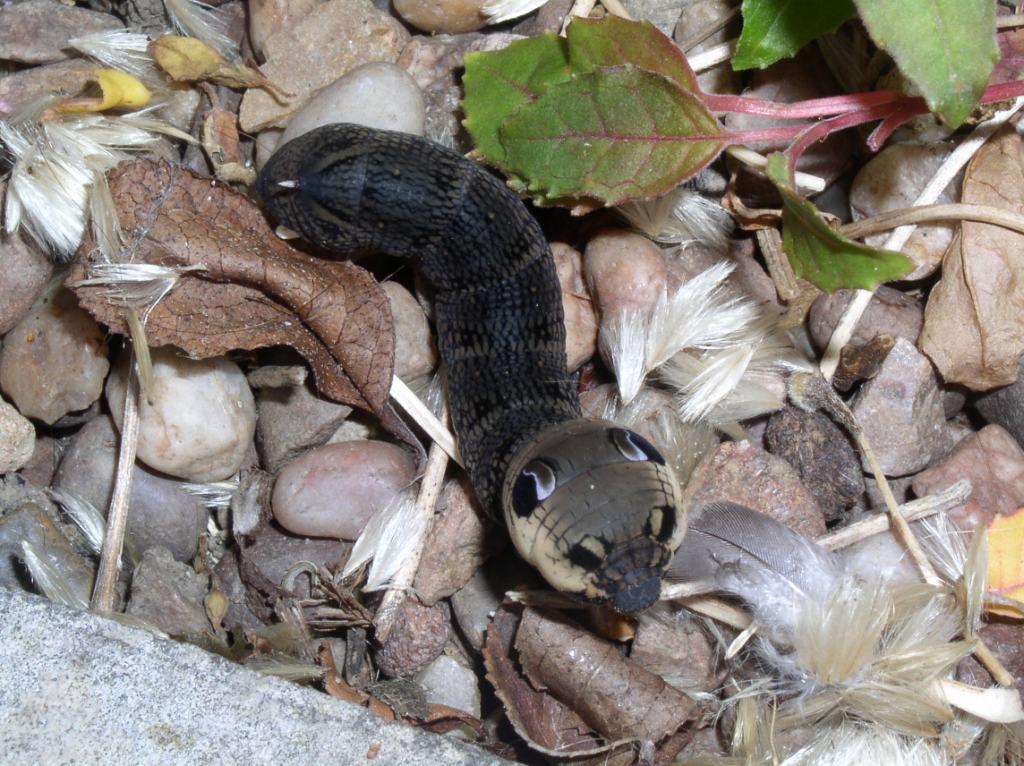
Mimicry using eyespots is widespread in nature - they are found in fish (such as the four-eyed butterflyfish Chaetodon capistratus which has them on the tail, so predators attack a non-lethal area or miss entirely), mammals (not only the 'obvious' ones such as leopards, but also the serval Leptailurus serval which has them on the backs of its ears for signalling to kittens while hunting), reptiles, birds and insects. Within the insects, butterflies and moths are probably best-known - many adult butterflies and moths have eyespots on the wings (the result of concentric pigment location around morphogenetic focus points), while the larva of the elephant hawkmoth Deilephila elpenor is famous for its conspicuous eye-like spots towards the head which are used to startle predators such as birds.

Elephant hawkmoth larva Deilephila elpenor showing eyespots
However, there are other invertebrates that show evidence of eyespots. I've previously written about bug (Hemiptera) nymphs possibly mimicing harvestmen, and today I noticed another - the common European garden spider Araneus diadematus. This is a very familiar species, often found on its orb-shaped web in gardens, and known for the pale cross-shaped marking (made from guanine which is a by-product of its protein metabolism) on the normally yellowish, orange or brown background of the top of the bulbous abdomen in females. Other common names include 'cross spider' and 'cross orbweaver', and males are smaller and less striking, though the markings are broadly similar.

Female Araneus diadematus showing the typical abdominal colour and cross-shaped marking

Male Araneus diadematus
So far, so good - but what about the eyespots I've mentioned. Well, the spiders are generally found either in the middle of their webs as shown above or tucked away in refuges at the ends of suspension silk lines. On webs they are typically head down and seen side on, either dorsally or ventrally. However, if you look stright down from the rear, a different pattern can be seen.

Female Araneus diadematus showing abdominal eyespots
To me, this is clear eyespot mimicry and makes adaptive sense - usually being head down, the rear of the abdomen is the part most likely to be presented to potential predators, namely birds, and therefore where eyespots that could startle them would be most useful. What I find more surprising is that I've never noticed this before despite having seen many specimens; more so that I can't find any other reports which suggests no-one else has either (or at least they written about it on the 'net). As ever, comments welcome!

Tuesday, 24 January 2012

Observations of Macleay's Spectres II: the boys

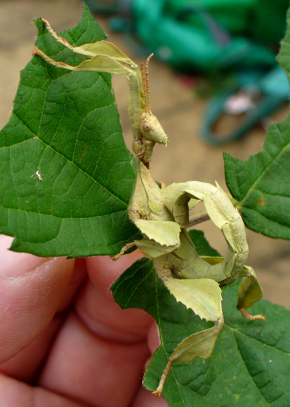
Having looked at the early stages (eggs and small nymphs) of my Macleay's Spectre stick insects (Extatosoma tiaratum), it's time to move on to looking at the older males. As they develop through a series of nymphal stages (instars), moulting and expanding in order to grow, they develop small spines and flanges, but most obviously a pair of wing buds. In their final (5th) instar, these are quite obvious and when they moult, emerging as an adult, these develop into full pleated wings with long 'coat-tail' covers. 

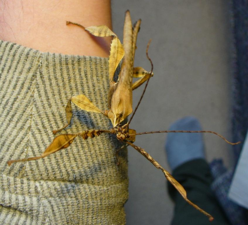
An adult male showing its long wing covers.
An adult male opening its wings just as it is about to take flight.
Once they have dried their wings (much like butterflies and other winged insects do), the males are able to fly strongly, and in the wild do so in order to find food and females, or to evade predation. However, I found that initially they were not very active apart from showing increased aggression when handled. During their final moult they also develop longer, curved antennae and larger, more protruding eyes, presumably used to find females both by scent at a distance and then visually when nearer. The wings fold along radial pleats (a bit like a parasol) and have a spotty pattern, and the neck is long and flexible at the joint with the thorax.

Head-on shot of a male in a threat posture showing well developed eyes and antennae.
The males remain well camouflaged, moving in a similar manner to leaves in the wind. For a few weeks they retained their variable colouring - some males were greenish, others brown or greyish. However, after this time a change seemed to take place with most darkening to a reddish-brown colour, and becoming more willing to fly - most evenings, the vivaria are opened and some males readily walk onto my outstreched hand and then launch (this is preceded by a subtle but definitie raising of the body into a launch posture), often having climbed onto my head or shoulder first. Around the same time, the first males could be found mating with the large adult females (more about them in Part III). So, it appears - albeit anecdotally - that this change in colour signals sexual maturity. If so, this is interesting as it does not seem to be associated with increased aggression either towards me (if anything they seem less aggressive when handled and simply fly more readily, though they may simply have habituated to handling, and some definitely avoid handling if possible) or other males. A number of males can be seen clustered around a female on occasions, but I have witnessed to overt aggression, though I have to assume that there is some form of competition to mate - maybe I need to observe what they are doing at 3am...

A male showing a dark red-brown colour and the long neck. The bright dot on top of the head is one of the male's ocelli (simple eyes).
A yellow-brown late (5th?) instar male nymph with the long wing-buds just visible behind the right middle leg.
Another late-instar (again, 5th?) male nymph, this one pale green in colour, again with the wing-buds visible.
When mating (or preparing to do so), males lie lengthwise along the back of a female (in the usual legs-outstretched 'stick' position) and both have genitalia at the rear of the flexible abdomens which bend to fit. The function of the long male neck with flexible articulation then becomes evident; females often arch backwards when feeding or moving and this forces the male's head backwards - the flexible neck allows him to remain in position without damage.

Males using my wife as a climbing-frame/launchpad while their cage is being cleaned. Just prior to this photo being taken, one of the males appeared to be trying to mate with her hair-grip (it has strong legs and a handy, accommodating central groove...)

Sperm transfer takes place in the form of a spermatophore - a packet of sperm in a hard 'shell' which novice insect keepers sometimes mistake for eggs. The spermatophore of E. tiaratum was noted by Clark (1975) and has been well documented since, but it was not until relatively recently that review of research and observations (e.g. Bragg, 1991) concluded that this structure provided the usual method of sperm transfer in the order Phasmida (AKA Phasmatodea). I recently collected an E. tiaratum spermatophore from the floor of one of my containers at home. The photo below shows the outer structure - the thread attaches it to the male during transfer to the female and the sperm-containing sac is 2.5-3mm in diameter, the whole being white with a pink tinge especially where the thread attaches to the sac.

Spermatophore of E. tiaratum
That's all for the males, but why not check out Part III: the girls...

References

Bragg, P.E. (1991). Spermatophores in Phasmida. Entomologist 110(2): 76‑80.
Clark, J.T. (1975). A conspicuous spermatophore in the phasmid Extatosoma tiaratum Macleay. Entomologist's Monthly Magazine 110: 81-82

Wednesday, 7 September 2011

Spider crabs - spiny substrates on legs

We're all familiar to some extent with crabs and other crustaceans - the shell or carapace, the pointed walking legs, the first pair of legs with pincers and so on. Around the (mainly southern and western) coast of Britain, one large and distinctive species is the Spiny Spider Crab Maja squinado which can have a carapace up to 20cm long (not to mention the long legs), although the specimen below is about half this size. This is a fairly shallow-water species, found sublittorally to about 50m depth, but sometimes also in deep littoral pools low on the shore.
Dorsal view of the carapace showing bumps (tubercles) and spines, including the two larger frontal spines of the rostrum seen spreading apart at the top of the photo. These rostral spines are straight unlike those of M. verrucosa which are curved outwards. Note the growths of other organisms such as seaweeds, hydroids and sponges.
The spines have clear potential as protection against predators, and can also prove uncomfortable to the unwary paddler if a carapace is trodden on (fortunately they are not poisonous like some organisms such as weaver fish) and indeed may hide under loose substrates such as coarse sand. Their ability to hide is enhanced by the range of other organisms that use them as a substrate to grow on e.g. sponges, hydroids and seaweeds - this allows them to blend in with other substrates supporting similar organisms. For those of you who like fairy tales, think about Baba Yaga's walking hut from the traditional Russian story, or indeed the Studio Ghibli animation Howl's Moving Castle!
A close-up of the upper surface of the carapace.
Zooming in on the carapace, the tubercles can be seen to have tufts of bristles - a close look shows that where some have broken off, tiny holes remain in the surface of the carapace. Such bristles or hairs are also seen on the legs following a moult, though these are gradually rubbed off. More of these bristles later...

The inner surface of the carapace.
In contrast to the patchily pinkish, rough outer surface (could this texture be to aid the attachment of organisms?), the inner surface, with no need to be involved in camouflage, is smooth and white. The dimples show that the tubercles are hollow, reducing weight (even so, with their spindly limbs and large bodies, large spider crabs can't support their own weight out of water) while the wrinkled arc shows where muscles and other tissues were attached when the animal was alive.

Another view of the underside; this time some of the points of attachment for the mouthparts and related appendages. The basal segment of one such appendage remains - its pair shows an empty socket.
A close-up of one of the articulations on the underside of the carapace.
The first of these two photos shows the membrane forming part of the joint at the base of the appendage, but some hairs are also visible. The second photo shows such a brush of hairs in more detail. They are simple unjointed structures and are likely to be guard hairs with the function of keeping sand grains and other unwanted material out of delicate joints.
The V-shaped notch between the two rostral spines showing numerous bristles as well as the tiny holes where some are missing.
The spaces between spines around the edge of the carapace show similar bristles. These may again be there to trap unwanted material (and maybe they help with camouflage by breaking up the outline, especially if they collect seaweed fragments and similar), but the crab also has much tinier, finer sensory hairs (not visible here) which also need to be protected by guard hairs. Of course, crabs do not rely solely on sensory hairs - they also have compound eyes on mobile stalks.
One mottled compound eye on its mobile stalk. This mobility means the eye can be tucked away into a notch in the carapace to prevent damage.

A close-up of the eye showing individual lenses and the 'seal' around the edge. Red and green iridescence is also visible just below the centre of the photo.

The base of the eye-stalk again showing the guard hairs which help to protect joints from damage by unwanted materials.
This has been a fairly brief tour of spider crab morphology, even though it covers only the carapace - if I find some during a bout of beach-coming, I may write a follow-up covering legs and other appendages. Until then, enjoy the story so far and we'll see what the tide and strandline bring...


References

Crothers, J. & Crothers, M. (1988). A key to the crabs and crab-like animals of British inshore waters. Field Studies 5: 753-806. Revised version available here.
Gibson, R., Hextall, B. & Rogers, A. (2001). Photographic Guide to the Sea & Shore Life of Britain & North-west Europe. OUP, Oxford.
Hayward, P.J. & Ryland, J.S. (eds) (2000). Handbook of the Marine Fauna of North-West Europe (reprint with corrections). OUP, Oxford.

Wednesday, 17 August 2011

Faking stinkers: Do bug nymphs mimic harvestmen?

A fairly short (well, short-ish) post for once, but one that asks a question that's been niggling at me for a while - do bug nymphs mimic harvestmen? At first glance this is not a question with a clear foundation, and certainly falls within the realm of 'speculative biology' (a place I don't visit that often) - bug nymphs don't look anything like harvestmen which have long spindly legs attached to a central round-oval body. However, by 'bugs' I'm only thinking of the Coreidae ('squashbugs') and Pentatomoidea ('shieldbugs'), and only one aspect of harvestmen (Opiliones) - the ocularium (eye-bearing structure) on top of the body. Let's look a little more closely...
A 'typical' harvestman of the genus Mitopus.

A North American harvestman, courtesy of 'bugman' at What's That Bug?

Both of these show the oval body on top of which is the small ocularium - a raised structure with two laterally oriented eyes which incidentally, despite appearing large like spider eyes, are ocelli and do little more than register light-intensity - touch is a far more important sense in harvestmen (Hillyard 2005). The top picture also shows the central dark band or 'saddle'. OK, so we know what harvestmen look like - but what's this got to do with bug nymphs? Well, let's see...

Late-instar nymph of the coreid Coriomerus denticulatus
Late-instar nymph of the coreid Arenocoris falleni
Final-instar nymph of the Juniper Shieldbug Cyphostethus tristriatus
[All three bug photos are copyright Tristan Bantock at the excellent British Bugs guide to British Hemiptera which has an extensive gallery including other nymphs showing these markings, though not all species do. For example, nymphs of the familiar Dock Bug Coreus marginatus do not show these 'ocularium' markings.]

Looking at these three nymphs, I can't help but wonder if the structures on the top of the abdomen are mimicing the ocalarium of harvestmen. To me, they certainly appear similar, especially in the coreids which share similar colouration and segmentation (superficially at least) with the harvestmen, though of course my poor human brain is wired to look for patterns, so simple appearance may not tell me much. I can do little more than speculate at present, and am unaware of anyone else who has looked at this, but the phrase 'Batesian mimicry' leaps to mind. This is the type of mimicry where a harmless species mimics one that is dangerous or unpalatable (for example, there are many species which mimic the colours of wasps). How might this suggested example be Batesian?

Well, bugs are likely to be potential prey of many insectivores, and during their nymphal stages cannot fly - so, mimicry might be useful, but why harvestmen? Although predatory, mainly on small soft-bodies invertebrates, harvestmen have no venom and are unlikely to be able to inflict much damage on potential predators such as birds, spiders, beetles, centipedes, fish, frogs and shrews. However, they do possess odoriferous glands (also called 'repugnatorial' or 'stink' glands) which are found on the sides of the carapace, approximately level with the ocularium. These glands produce a spray or droplet than the harvestman can spread on itself or an attacker. Though the chemicals (various alcohols, ketones and naphthoquinones) are not always easily detected by humans' sense of smell, there have been various observations made of potential predators avoiding (e.g. ants and spiders) or expelling (e.g. frogs) harvestmen because of their distastefulness (Hillyard 2005). Might this form the basis of mimicry by bug nymphs?

At present this is mere speculation derived from observation - something that might be formulated as a hypothesis - and some initial questions are raised such as whether the bug nymphs themselves are distasteful, in which case this would be an example of Müllerian, rather than Batesian, mimicry (as both groups have anti-predation characteristics). It is something that I will have to dig into further and as ever I welcome thoughts and suggestions.

Reference

Hillyard, P.D. (2005). Harvestmen (3rd ed.). Field Studies Council, Preston Montford. A small but excellent book, and the current standard work on British harvestmen.

Further reading

If you are interested in squash- and shieldbugs in Britain, this is excellent:

Evans, M. & Edmondson, R. (2005). A Photographic Guide to the Shieldbugs and Squashbugs of the British Isles. WGUK.