Welcome

Welcome to my blog

This is where I post various musings about wildlife and ecology, observations of interesting species (often invertebrates)
and bits of research that grab my attention. As well as blogging, I undertake professional ecological & wildlife surveys
covering invertebrates, plants, birds, reptiles, amphibians and some mammals, plus habitat assessment and management
advice
. I don't work on planning applications/for developers. The pages on the right will tell you more about my work,
main interests and key projects, and you can follow my academic work here.
Showing posts with label moss. Show all posts

Monday, 25 February 2013

Eight spores good - a mossy mystery

Looking through moss and fragments of moist dead wood recently, I've found a range of small invertebrates such as mites, and spurred on to see what else dwells within, I've clocked up some more microscope time. Scanning a specimen of the creeping feather-moss Amblystegium serpens, a common species known from various habitats/substrates including both living and dead wood, I noticed some tiny red-brown structures underneath some of the leaves. Having a look through Atherton et al. (2010) and Watson (1981), no moss structures looked quite like it, so I started photographing and magnifying...

Structures growing from the stem of Amblystegium serpens
There's not a lot of detail here, but an idea of scale can be gained - the width of the stem (no more than about 0.2mm - the leaves are also tiny, about 0.5mm long) is clear where it reaches the left-hand side of the photo and the more-or-less rectangular (but not especially elongate) individual cells can be seen there. Just to the left of the pin, the stem is a little different however - the cells are orange-brown and a little larger. This might be of interest as many gall-causing Fungi induce changes in cell size and tissue colour. So, to see more detail in the brown masses, I made a simple 'squash' preparation for higher magnification.

Fibrous structure growing from the stem beneath a leaf of Amblystegium serpens
The fibrous structure is clearer here, including its point of attachment/outgrowth from just below the base of a leaf. The longest fibres are maybe 0.5mm long, maybe a little more, but it is still unclear exactly what they are. Mosses produce a number of structures worth considering here:

  • Paraphyses - thin sterile hairs, sometimes club-shaped, usually multicellular. A possibility.
  • Protonema - the young stage of a moss that develops when a spore germinates; generally appears as a system of green threads. Clearly not the case here.
  • Gemma - a unit of vegetative propagation, may be single-celled, two-celled or multicellular. Another possibility.
To determine whether these were parts of the moss and/or a fungus growing on it, more detail was needed.

Various structures seen attached to Amblystegium serpens
A fungal spore found with a sample of Amblystegium serpens
In the first of this pair of photos, a number of structures are visible. Those indicated by blue arrows are unidentified - the larger fragment could be a paraphysis or similar (or an equivalent fungal paraphysis) while the 4-celled structure could be gemmal or a 4-celled fungal ascus (spore-bearing 'sac') - this is largely guesswork however, partly informed by checking fungal structure using Webster (1970). The structure indicated by a red arrow is rather clearer and appears to be the ascus (spore-bearing 'sac') of a discomycete fungus, complete with the typical eight spores seen when fully developed, though the left-most one is blurred out in the photo. Other similar structures could be seen, including the spore in the lower photo - it is almost spherical and measures around 13 x 15 um, and although again blurred out here, has a surface covered in tiny 'warts' (i.e. it is 'verruculose'). Consulting Ellis & Ellis (1998), there appears to be only one contender with this host and set of characteristics - the microfungus Octospora wrightii which is associated primarily with this moss, and is found from January to March.


As ever, comments, suggestions and corrections welcome - I am, as is so often the case, writing outside my comfort zone here; how else to learn though?


References

Atherton, I., Bosnaquet, S. & Lawley, M. (eds.) (2010). Mosses and Liverworts of Britain and Ireland: A Field Guide. British Bryological Society. [If you only buy one UK bryology book, I can recommend making it this one]
Ellis, M.B. & Ellis, J.P. (1998). Microfungi on Miscellaneous Substrates: An Identification Handbook (2nd ed.). Richmond, Slough.
Watson, E.V. (1981). British Mosses and Liverworts (3rd ed.). Cambridge University Press.
Webster, J. (1970). Introduction to Fungi. Cambridge University Press.

Wednesday, 20 February 2013

Sighting the mighty tiny mite

A couple of days ago, I mentioned oribatid mites, more-or-less in passing while looking at other moss, soil and decay fauna. As they are so important in the processes of decay and decomposition, feeding on all sorts of organic matter, I decided to return to my samples and see what else I could find. Unsurprisingly I found a few more but although I can't identify them to species (yet - I'm starting to have a look at Michael (1888) which you can download for free here), I thought it would be a good opportunity to give a bit of introductory information about this often unfamiliar group. So, what are oribatids?

Taxonomically, they form the order Oribatida within the Acari (mites) which are in turn arachnids. All are small (none more than 1.4mm in length) and those that I found were between 0.5 and 1.0mm. They are largely found in soils (especially in woodlands) and decaying matter (dead wood, leaf litter etc), and are important in soil processing and formation in much the same way as earthworms. Like other arachnids they have jaws known as chelicerae, and are eight-legged, but some other features may not be familiar.

Oribatid mite, approx 0.7mm long, ventral view
The pteromorph is a wing-or cloak-like extension of the carapace which wraps partly around the mite, presumably to armour it against damage from soil particles or predators. The genital shield is a round structure formed of two semi-circular halves which covers the reproductive structures - males have a penis-like structure called an aedeagus, much like that seen in beetles. Similarly, the anal shield is paired and covers the end of the gut.

Oribatid mite, approx 0.7mm long, ventral view
Unlike the more familar red spider mites (Trombidium spp.), these are not hairy/velvety but are smooth with only fine sculpturing and sometimes larger but fewer bristles. The legs do however have long bristles which perform a sensory function, and in fact the first pair of legs of some species are purely sensory (like antennae) and are no longer used for locomotion. There is a single median eye and with vision reduced (unsurprisingly as oribatids are mainly soil-dwelling), sensory bristles have become more important, not just on the legs, but also via the development of specialised bristles to form a 'pseudostigmatic organ' which can have a variety of shapes e.g. club or drumstick.

That is all the detail I can provide at present without simply summarising Michael (1888) and covering features not visible in my photographs. However, I am sure oribatids will show up on my invertebrate radar again, and I hope to be able to delve more deeply into their ecology and taxonomy.

Reference

Michael, A.D. (1888). British Oribatidae Vol.II. Ray Society, London.

Monday, 18 February 2013

Tiny denizens of the rot-hole

What with winter keeping most invertebrates out of sight, it's been a while since I wrote much of an entomological nature, but yesterday was a fine opportunity to head up to Beacon Hill nature reserve to see what was about. As well as chalk grassland (which will be more interesting when in flower), there is an interesting stand of beech woodland and associated scrub, including an ecologically important resource of dead wood (standing and fallen) and old mossy trees.

The base of a mossy beech tree.
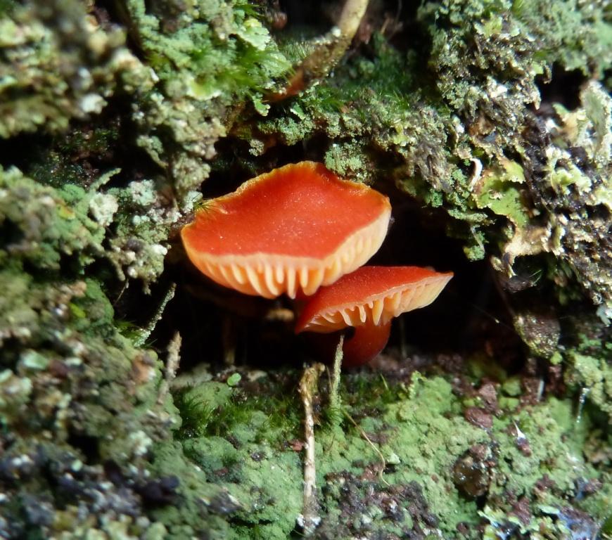
These features mean that there is habitat for many fungi and dead-wood invertebrates and the evidence is everywhere - beetle boreholes (some opened by woodpeckers, one of which could be heard clearly in the woodland), wood in various stages of decay, insect-feeding birds such as a treecreeper (Certhia familiaris) and an intriguing-looking rot-hole with a large fungus and a tuft of hair poking out of it...

Rot-hole with fungus and tuft of hair
Looking inside, it had been lined with moss and hair, and was clearly a nest or roost of some sort, either of a bird or small mammal, and the fungus is (I think) an oyster mushroom Pleurotus ostreatus - if any mycologists would like to correct me on this, please do!

The inside of the rot-hole; at the base of the fungus, a layer of moss-and-hair bedding.
So, sample-pot in hand (like any good ecologist!), I took a pinch of the mossy bedding, including some soil/decayed wood from directly beneath it - after all, there are plenty of under-recorded parasites that live in vertebrate nests and you never know what you'll find. Back home, it was time to check what I'd found; some of the small inveretebrates such as Collembola (springtails) hide very effectively in material like this, so I find that adding some water to the sample in a watch-glass causes them to float to the surface and become easier to see. Indeed, doing this brought up a cluster of the common springtail Ceratophysella bengtssoni which can sometimes be found in large aggregations on the surface of soil and puddles, but may well still have been hibernating given the cold night-time temperatures at present.

Several Ceratophysella bengtssoni from the nest sample
These weren't the only springtails - Lepidocyrtus cyaneus and Neanura muscorum were also present, as was Tomocerus vulgaris from mossy dead-wood of a nearby tree. The samples included quite a few empty moulted skins, suggesting that these are not solely hibernation sites, but places where active feeding and growth occur - unsurprising as they mainly feed on fungal hyphae and decaying plant material (no shortage in this sample location). They are also an excellent group to look for in the winter as they can be found throughout the year - if you are interested in the UK species, Hopkin (2007) is an excellent place to start. However, Collembola are not the only soil/leaf-litter animals to be found. Hidden among the tangle of hair (mainly sheep I think) and plant fibres were two shed skins of an oribatid soil mite.

The shed skin of an oribatid soil mite
Oribatids are beyond my identification skills (I don't even know anyone who can ID them, though I do have a go at halacarids occasionally), but the shiny bulbous shape, the pointed mouthparts and the leg attachment points are all visible here. Though poorly known outside the realm of specialists, these mites are important in the decay process, feeding on a wide range of plant, animal and fungal organic material, with a minority being predatory - in fact they break down and process soil material in a similar way to earthworms even if they aren't as familiar or well-understood/studied.

Another species, common if often over-looked, and mainly found under bark or logs in woodland is the spotted snake millipede 
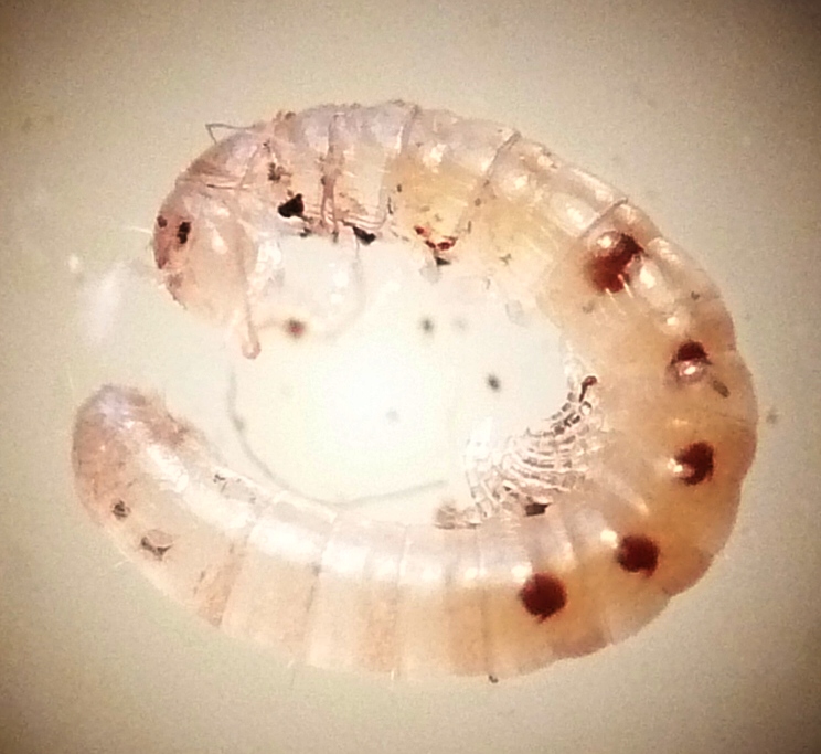
Blaniulus guttulatus. It is often considred a pest (e.g. in allotments) but probably only enters crops when damage has already occurred, such as by a 'primary' pest or some other mechanical means. They grow to around 20mm in length and are white with rows of red spots along the sides. The specimen I found however was a juvenile no more than about 3mm long (with few segments/spots), and the first early stage I've seen of this species.

Juvenile Blaniulus guttulatus
So, a few interesting finds - common species (no idea about the oribatid) but indicative of the small and often un-noticed soil/dead-wood fauna. Interestingly there were no mammal/bird nest-dwelling species (such as ticks or fleas), and no indication of exactly what had been using the hole - however, the presence of fine hairs and small dark elongate faeces, plus a lack of even small feathers suggest a mammal, presumably a rodent, rather than a bird.

Reference

Hopkin, S.P. (2007). A Key to the Collembola (Springtails) of Britain and Ireland. FSC, Shrewsbury.

Monday, 26 November 2012

What's in a wall?

As you might imagine, I don't mean bricks and mortar - instead I am more interested in the species that can be found growing on walls, particularly old rough stone walls in areas with low levels of air pollution such as might be found in the village of Slapton in south Devon. Yup, where I was one holiday a couple of weeks ago... Old walls are well known to be important for wildlife - for instance, lizards can often be seen basking on them - but for those of us living or working in cities, especially with relatively new and smooth-surfaced buildings (and more air pollution), the opportunities to see such species can be limited.

Some are so strongly associated with this type of habitat that their common names reflect it, such as the wall pennywort Umbilicus rupestris (the generic name reflects its other common name, 'navelwort'). Like many wall-dwelling species, they are also found on natural rocks, but man-made rocky surfaces can be just as good.

The round leaves of wall pennywort Umbilicus rupestris.
You can see pale green lichenous growth here, but a closer look around this wall revealed a somewhat more impressive display - the little trumpet-like fruiting bodies of the lichen Cladonia (one of several similar species).
A cluster of Cladonia
There were also larger growths of non-lichenous fungi among the damp, mossy hollows. One was the branched and spindly Grey Coral Clavulina cinerea. This is a common species but being associated with the ground in woodlands, might not seem a likely wall-colonist. However, the crevices were full of moss and soil, and roots were present from the woody plants on top of the bank the wall retains. So, from the fungus' point of view, just like home! Another was an attractive orange-red waxcap Hygrocybe sp., possibly H. helobia which was only described scientifically in 1974 and is more often associated with grassy woodland clearing or heaths (but again, conditions are suitable in this wall). However, without closer examination (beyond what I'm likely to do on holiday), the species-level ID remains tentative.
Grey Coral fungus Clavulina cinerea

Waxcap Hygrocybe sp.
Moving through the major taxonomic groups, there are also ferns associated with walls. One of these is the rustyback Asplenium ceterach which I initially thought I had found; however, closer examination (yes, sometimes even on holiday, though it took iSpot to confirm it as I don't ID ferns very often...) showed it to be A. adiantum-nigrum, though both are common in SW England. Another (which I can also find growing from crevices in the walls of the church near where I live) is the maidenhair spleenwort A trichomanes.
Asplenium adiantum-nigrum (upper surface of fronds)

Asplenium adiantum-nigrum (lower surface of frond showing red-brown sori)

Maidenhair spleenwort Adiantum trichomanes
Lastly I want to return to the flowering plants, this time a non-native garden escape Nerine bowdenii, also known as the Guernsey Lily. This southern African species is a popular plant in the village of Slapton and can be seen along the narrow lanes outside many of the cottages. However, coming from mountainous areas, it too can adapt to 'mural' life and at least one had managed to escape through a garden fence and grow on top of the old stone wall featured here. I could continue - after all, with all these plants, fungi and lichens, there are of course many invertebrates (my usual topic!) - however, as I promised my wife I would not spend the week rummaging around with collecting pots, these will remain mysterious for now and I'll simply leave you with the big pink lily which, although appearing delicate, was clearly capable of surviving repeated buffeting by passing vehicles...
Guernsey Lily Nerine bowdenii.

Monday, 16 January 2012

Teeny-tiny toadstools - or are they?

Yesterday I was pointed towards a log cut from a fallen tree in my in-laws' garden. On it was a fairly dried-out growth of moss and on the leaves, what looked like some tiny (1mm) fungal caps. Now, I'm no mycologist (or bryologist), but I do find microfungi intriguing and happen to have a copy of Ellis & Ellis (1998) which is the standard (only?) work covering British microfungi on substrates other than vascular plants - for those you need Ellis & Ellis (1997).

The specimen - the caps are about 1mm in diameter.
A slightly closer view showing the wrinkled stalks.
The moss is fairly dry and a bit of a miserable specimen without capsules, but looking at the excellent Atherton et al. (2010), I think that it is the common and widespread species Brachythecium rutabulum (sometimes known as the Rough-stalked Feather-moss). The fungus itself is white with a split cup-like cap and a wrinkled stem that widens towards the base. The cap is granular beneath and bears hairs in the upper/outer surface.
Two of the fruiting bodies showing details of stems and caps.
Side view of a fruiting body showing the split cap and granular spore-bearing structures.
Some moss-epiphytic microfungi have clear fringes of hairs around the edge of the cap. This doesn't appear to be the case here - instead there appears to be an irregular tufting of hairs on the top of the cap with a few at the edge (I wondered if they were the hyphae of another even smaller fungus, but I don't think so).

Side view of the cap showing tufts of hairs.

High-power image of the cap showing branching hairs.

Lastly, spores are an important feature used in the identification of microfungi. I collected some of these on a slide and they are clearly almost spherical and reddish-brown in colour, and each is about 10um in diameter.

Spores (x100 magnification)

So, the overall size, colour, spore colour/shape/size, habitat, hairs and other features lead me to tentatively identify this as Chromocyphella muscicola, a species with caps up to about 3mm diameter which is associated with mosses on bark. However, having consulted with a mycologist from the Hampshire Fungus Recording Group, it appears that C. muscicola doesn't produce stalks like this. Other superficially similar genera such as Leptoglossum also differ from this specimen in one or more ways e.g. spore shape, cap hairs. And so, it was suggested that this might not be a true fungus at all, but instead a myxomycete or 'slime mould' where the globular sporangium had broken open to reveal the network of spore-bearing hairs known as the capillitium.

I have posted about myxomycetes before (e.g. here and here) but it still a group of organisms I know little about, fascinating as they are - taxonomically protozoans but traditionally treated as 'honorary' Fungi. So, swapping books to look at Ing (1999), it soon became clear that this was indeed a 'myxo'. It also happened to be one that was relatively easy to identify from the keys and descriptions - the pale colour, size, stalk, open split 'cap' with the hairs of the capillitium, and spores (shape, size, colour, texture). This process also taught me that an important identification feature can be the shapes, colours and sizes of nodes of the capillitium i.e. structures about the size of a spore where hairs meet and branch.

These features all combined to indicate that this is the myxomycete Physarum nutans - a common and widespread species, though rarely found on mosses (it is usually on dead wood or the bark of live trees). It is very similar in appearance to Didymium squamulosum but they can be separated by features such as the spores (in D. squamulosum they are dark brown in transmitted light whereas in P. nutans, as here, they are pale brown). So, an interesting process for me, looking at an unfamiliar group - the only way to learn more about species identification!

References

Atherton, I., Bosanquet, S. & Lawley, M. (eds.) (2010). Mosses and Liverworts of Britain and Ireland: A Field Guide. British Bryological Society.
Ellis, M.B. & Ellis, J.P. (1997). Microfungi on Land Plants: An Identification Handbook. Richmond, Slough.
Ellis, M.B. & Ellis, J.P. (1998). Microfungi on Miscellaneous Substrates: An Identification Handbook. Richmond, Slough.
Ing, B. (1999). The Myxomycetes of Britain and Ireland: An Identification Handbook. Richmond, Slough.