Welcome

Welcome to my blog

This is where I post various musings about wildlife and ecology, observations of interesting species (often invertebrates)
and bits of research that grab my attention. As well as blogging, I undertake professional ecological & wildlife surveys
covering invertebrates, plants, birds, reptiles, amphibians and some mammals, plus habitat assessment and management
advice
. I don't work on planning applications/for developers. The pages on the right will tell you more about my work,
main interests and key projects, and you can follow my academic work here.
Showing posts with label mimicry. Show all posts

Wednesday, 19 June 2013

Murk-dwelling bottom-breathers

I've posted about our garden pond before, but may not have mentioned that off to one side of it is an old stone fountain-top which has been plugged to provent leaking. Having no outflow unless there is heavy rain and it spills over, this means it becomes stagnant with leaves and other organic matter accumulating. The temptation might be to clean this out and add clean water, but no, it is there for a reason - habitat for larvae that are adapted for such conditions. An example of this is the rat-tailed maggot, the larva of hoverflies in the genus Eristalis, in particular E. tenax, and others such as the sun-flies Helophilus sp., plus Sericomyia, Mallota, Anasimyia and Myathropa. As adults, Eristalis are are excellent honey-bee mimics (hence the common name of 'drone-flies'), but their larvae, like those of the other genera listed, are very different. The most obvious feature is the long, telescopic posterior breathing tube - essential when living in stagnant, low-oxygen water among decaying vegetation.
Rat-tailed maggot, the larva of Eristalis sp. (probably E. tenax), approx. 10mm long excluding 'tail'.
This is a young larva - my identification is a little tentative as I can't see the key features yet, but the timing is right as E. tenax were frequenting the water long enough ago for eggs to now have hatched, whereas although there are Helophilus pendulus as well, they have only just started breeding activity as far as I can tell; I will be able to confirm their identity when they are more fully developed. They are filter-feeders and some of the gut contents are visible here, as are the well-developed prolegs.

Rat-tailed maggots showing how their breathing tubes are used.
The white section of the tube forms a sheath made from one extended segment and contains complex musculature used to extend and retract the breathing tube, as well as protecting it (the tip of the tube can be seen as a short white-tipped dark segment). The larvae can also swim slowly by undulating the body and tube. They may not be the most attractive creatures, but I do think they are interesting, and they are of evolutionary interest as these larval forms appeared quite recently in hoverfly evolutionary history (Rotheray, 1993), not to mention their role in consuming waste organic material. They do of course also develop into adults - the dronefly mentioned above, which has a role in pollination, so a valuable species too.

Eristalis adult on Buddleia davidii

Reference

Rotheray, G.E. (1993). Colour guide to hoverfly larvae (Diptera, Syrphidae). Dipterists Digest 9: 1-156.

Thursday, 13 September 2012

Eyes in the back of my... back...

Mimicry using eyespots is widespread in nature - they are found in fish (such as the four-eyed butterflyfish Chaetodon capistratus which has them on the tail, so predators attack a non-lethal area or miss entirely), mammals (not only the 'obvious' ones such as leopards, but also the serval Leptailurus serval which has them on the backs of its ears for signalling to kittens while hunting), reptiles, birds and insects. Within the insects, butterflies and moths are probably best-known - many adult butterflies and moths have eyespots on the wings (the result of concentric pigment location around morphogenetic focus points), while the larva of the elephant hawkmoth Deilephila elpenor is famous for its conspicuous eye-like spots towards the head which are used to startle predators such as birds.

Elephant hawkmoth larva Deilephila elpenor showing eyespots
However, there are other invertebrates that show evidence of eyespots. I've previously written about bug (Hemiptera) nymphs possibly mimicing harvestmen, and today I noticed another - the common European garden spider Araneus diadematus. This is a very familiar species, often found on its orb-shaped web in gardens, and known for the pale cross-shaped marking (made from guanine which is a by-product of its protein metabolism) on the normally yellowish, orange or brown background of the top of the bulbous abdomen in females. Other common names include 'cross spider' and 'cross orbweaver', and males are smaller and less striking, though the markings are broadly similar.

Female Araneus diadematus showing the typical abdominal colour and cross-shaped marking

Male Araneus diadematus
So far, so good - but what about the eyespots I've mentioned. Well, the spiders are generally found either in the middle of their webs as shown above or tucked away in refuges at the ends of suspension silk lines. On webs they are typically head down and seen side on, either dorsally or ventrally. However, if you look stright down from the rear, a different pattern can be seen.

Female Araneus diadematus showing abdominal eyespots
To me, this is clear eyespot mimicry and makes adaptive sense - usually being head down, the rear of the abdomen is the part most likely to be presented to potential predators, namely birds, and therefore where eyespots that could startle them would be most useful. What I find more surprising is that I've never noticed this before despite having seen many specimens; more so that I can't find any other reports which suggests no-one else has either (or at least they written about it on the 'net). As ever, comments welcome!

Thursday, 26 January 2012

Observations of Macleay's Spectres III: the girls

Following sections covering eggs and early nymphal stages and mature males, the third part of this series on Extatosoma tiaratum, the Macleay's Spectre, looks at mature females.

Female E. tiaratum with front legs raised.
As late-instar nymphs and adults, females are structurally very different from males (sexual dimorphism). They are larger - up to 150mm long with males around 100mm, broader and much heavier - up to around 30g (a lot for an insect and females often drag the spiny underside of their abdomen along the surface they are walking on, sometimes producing a clear scraping noise as they move). Females also have many more spines and flanges, including a more pronounced 'head-dress' of short blunt spines. In fact, in E. tiaratum, the differences between males and females are so pronounced that they were originally described as separate species (Hadlington & Johnston, 1998)! Details of female genitalia are given by Heather (1965) and note that from a sample of seven specimens there were on average 151 ovarioles (tubes forming the paired ovaries) with an average of seven oocytes (immature egg cells) each. The bursa copulatrix (the sac-like organ where the spermatophore or 'sperm packet' is stored after copulation) bears around 20 blind tubules and there appears to be no distinct spermatheca (sac for sperm storage) or other sclerotised (hardened) part of the genitalia. This matches the observation (see Part II) that sperm transfer is by spermatophore.

Flanges and spines on the leg of a female.
The spine-covered abdomen of a large female.
Side view of a female's head showing the oval extension of the top with its 'head-dress' of short spines. Also note that the camouflage includes marking breaking up the shape of the eye.
The head of a large female showing small wart-like bumps as well as a clear view of the mouthparts.
Around the same time my males changed to a brick-red colour, my two large females developed a pinkish-lilac tinge as shown in the photo of the abdomen above. This may be an indication of sexual maturity or possibly gravidity (containing eggs). Also, following their final moult, the adult females became noticeably more aggressive, making for interesting cage-cleaning sessions... This aggression included general attempts at evading capture (fleeing or dropping out of reach; previously these had not occurred often) plus active attempts to use spines as weaponry (such as pinching with the inner edge of a bent leg, nut-cracker style), including against unwary males that strayed too close soon after an angry female was replaced in the cage. I have seen one male with what appears to be a small healing puncture wound (i.e. a hardened drop of what I imagine to be dark haemolymph) on the ventral side of the thorax though whether this was caused by female aggression or an accident with a bramble thorn I do not know.

A female nymph already showing spines and flanges.
Side view of a late-instar female nymph.
As this pair of photos shows, the female structure develops at a relatively early stage, essentially just getting larger with each moult. This contrasts to some extent with males which show a number of significant structural changes following their final moult.

A big girl perching on my wrist. She is mighty!
So, I started this series of posts with eggs, and can return to this topic as two days ago one of the large females began laying eggs. This is not a precise process in E. tiaratum as eggs are flicked up to a few feet (maybe a metre or so) by twictching the abdomen when the egg is laid (this also occurs with frass AKA insect faeces which takes the form of dry 3mm x 5mm oval pellets of plant material - in males, frass is long and thin, approx 1mm x 5mm). Fortunately as my insects are in cages, the eggs are easy to collect so I hope to have another generation relatively soon.

Tip of a mature female's abdomen showing an egg about to be laid. Note the dorsal spines on the abdomen - they are splayed outwards which may be an adaptation to accommodate a male during mating.

A female nymph doing her favourite thing - eating bramble leaves, nom nom nom.

It's busy during cage-cleaning time.

References

Hadlington, P & Johnston, J.A. (1998). An Introduction to Australian Insects (revised ed.). University of New South Wales, Sydney.
Heather, N.W. (1965). Studies on female genitalia of Queensland Phasmida. Australian Journal of Entomology 4(1): 33-38.

Tuesday, 24 January 2012

Observations of Macleay's Spectres II: the boys

Having looked at the early stages (eggs and small nymphs) of my Macleay's Spectre stick insects (Extatosoma tiaratum), it's time to move on to looking at the older males. As they develop through a series of nymphal stages (instars), moulting and expanding in order to grow, they develop small spines and flanges, but most obviously a pair of wing buds. In their final (5th) instar, these are quite obvious and when they moult, emerging as an adult, these develop into full pleated wings with long 'coat-tail' covers. 

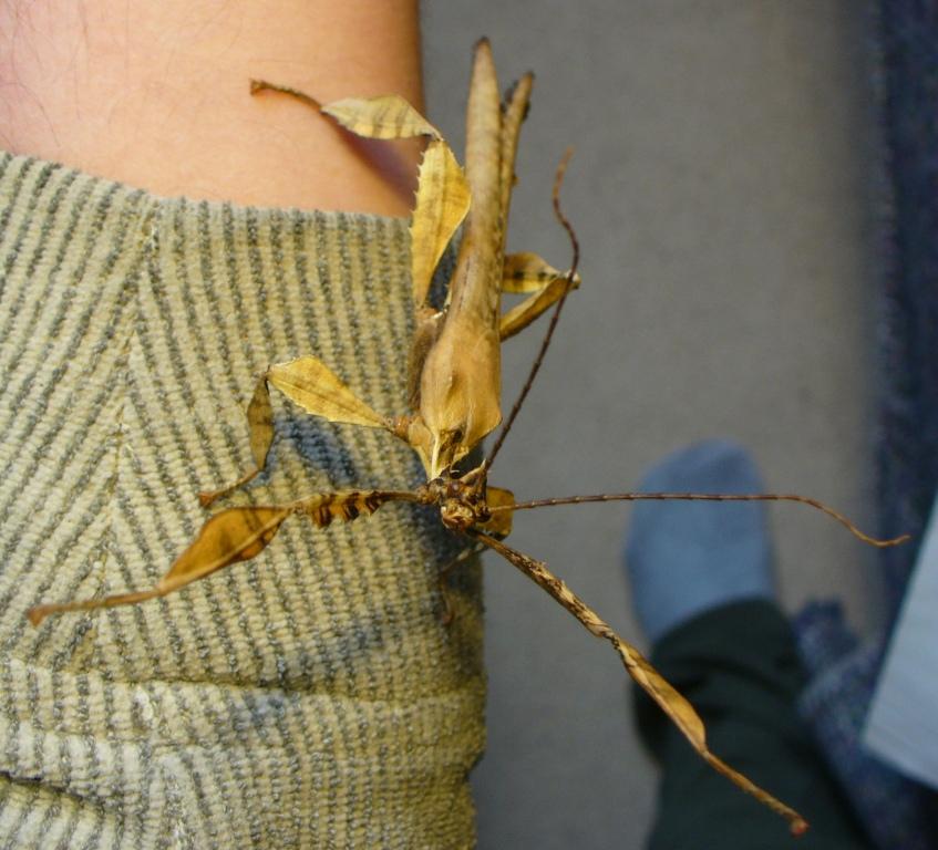
An adult male showing its long wing covers.
An adult male opening its wings just as it is about to take flight.
Once they have dried their wings (much like butterflies and other winged insects do), the males are able to fly strongly, and in the wild do so in order to find food and females, or to evade predation. However, I found that initially they were not very active apart from showing increased aggression when handled. During their final moult they also develop longer, curved antennae and larger, more protruding eyes, presumably used to find females both by scent at a distance and then visually when nearer. The wings fold along radial pleats (a bit like a parasol) and have a spotty pattern, and the neck is long and flexible at the joint with the thorax.

Head-on shot of a male in a threat posture showing well developed eyes and antennae.
The males remain well camouflaged, moving in a similar manner to leaves in the wind. For a few weeks they retained their variable colouring - some males were greenish, others brown or greyish. However, after this time a change seemed to take place with most darkening to a reddish-brown colour, and becoming more willing to fly - most evenings, the vivaria are opened and some males readily walk onto my outstreched hand and then launch (this is preceded by a subtle but definitie raising of the body into a launch posture), often having climbed onto my head or shoulder first. Around the same time, the first males could be found mating with the large adult females (more about them in Part III). So, it appears - albeit anecdotally - that this change in colour signals sexual maturity. If so, this is interesting as it does not seem to be associated with increased aggression either towards me (if anything they seem less aggressive when handled and simply fly more readily, though they may simply have habituated to handling, and some definitely avoid handling if possible) or other males. A number of males can be seen clustered around a female on occasions, but I have witnessed to overt aggression, though I have to assume that there is some form of competition to mate - maybe I need to observe what they are doing at 3am...

A male showing a dark red-brown colour and the long neck. The bright dot on top of the head is one of the male's ocelli (simple eyes).
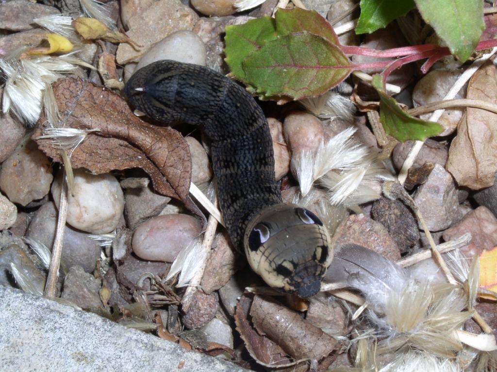
A yellow-brown late (5th?) instar male nymph with the long wing-buds just visible behind the right middle leg.
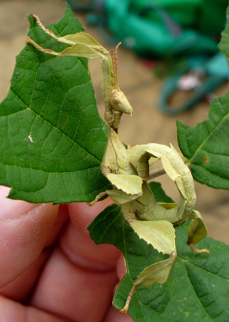
Another late-instar (again, 5th?) male nymph, this one pale green in colour, again with the wing-buds visible.
When mating (or preparing to do so), males lie lengthwise along the back of a female (in the usual legs-outstretched 'stick' position) and both have genitalia at the rear of the flexible abdomens which bend to fit. The function of the long male neck with flexible articulation then becomes evident; females often arch backwards when feeding or moving and this forces the male's head backwards - the flexible neck allows him to remain in position without damage.

Males using my wife as a climbing-frame/launchpad while their cage is being cleaned. Just prior to this photo being taken, one of the males appeared to be trying to mate with her hair-grip (it has strong legs and a handy, accommodating central groove...)

Sperm transfer takes place in the form of a spermatophore - a packet of sperm in a hard 'shell' which novice insect keepers sometimes mistake for eggs. The spermatophore of E. tiaratum was noted by Clark (1975) and has been well documented since, but it was not until relatively recently that review of research and observations (e.g. Bragg, 1991) concluded that this structure provided the usual method of sperm transfer in the order Phasmida (AKA Phasmatodea). I recently collected an E. tiaratum spermatophore from the floor of one of my containers at home. The photo below shows the outer structure - the thread attaches it to the male during transfer to the female and the sperm-containing sac is 2.5-3mm in diameter, the whole being white with a pink tinge especially where the thread attaches to the sac.

Spermatophore of E. tiaratum
That's all for the males, but why not check out Part III: the girls...

References

Bragg, P.E. (1991). Spermatophores in Phasmida. Entomologist 110(2): 76‑80.
Clark, J.T. (1975). A conspicuous spermatophore in the phasmid Extatosoma tiaratum Macleay. Entomologist's Monthly Magazine 110: 81-82

Sunday, 22 January 2012

Observations of Macleay's Spectres I: early stages

Last summer, I was given an unusual birthday present - a 35mm film canister full of eggs of the Macleay's Spectre stick-insect Extatosoma tiaratum, sometimes known as the Giant Prickly stick-insect. These are found mainly in the Australian forests of Queensland and New South Wales where they feed on the foliage of eucalyptus trees, although in captivity they eat a range of unrelated species - one popular food is bramble (Rubus fruticosus agg.) which is what I use as it is readily available for free. However, young bramble leaves can contain noxious chemicals that make them unpalatable or even toxic, especially to young insects - I tend to remove these, and when some have been included by accident with more palatable older leaves, the insects ignored them.There is plenty of information about this species (e.g. from the Phasmid Study Group) as they are popular pets for those who like their companion animals to be six-legged, but I have made some observations which may be of interest. So, let's start at the beginning - with eggs...

Egg of E. tiaratum with the lid (operculum) open.
Close-up of the operculum showing the translucent sealing membrane around the edge. There is another membrane below this which seals the opening to the egg and which is broken to allow emergence by the first-instar nymph.
The eggs are a few mm long, oval with a sculptured ridge along one long axis, and a round lid (operculum). They are speckled with various shades of yellow and brown, camouflaging them to look like seeds when they fall to the forest floor in the wild. In those I have at home, the eggs are a pale yellow-brown when freshly laid and dry, but when placed on damp tissue to (eventually) hatchm they become a dark brown - presumably this aids camouflage with dry soil conditions also being paler and darkening when there is rain. When females lay eggs, they often flick them several feet by twitching their abdomens, presumably to aid dispersal  - the function of this might be to prevent a cluster all being eaten in one go by a predator. The eggs have a coating (made of lipids and other organic materials) which is edible to ants and which induces them to take the eggs to their colony and eat this coating. The eggs remain otherwise intact and the ants dump them on the colony's waste pile where they hatch. Thus the eggs are likely to become clustered following the initial dispersal during laying but will have spent much of their time in an ant colony where the chance of predation is much reduced. Luckily, the eggs can hatch without the attention of ants which means that those missed by foraging ants and of course those in captivity remain viable.

Unsurprisingly, given their birthplace, E. tiaratum hatchlings (forst-instar nymphs) are ant-mimics, specifically of the large, long-legged 'spider ants' in the genus Leptomyrmex.

Recently hatched nymph showing the orange head and black body providing its mimicry of Leptomyrmex ants.
The orange head and black body, as well as overall shape closely mimics that of ant species such as L. darlingtoni, L. erythrocephalus and L. rufipes and so benefit from appearing similar to these toxic species (the sting has degenerated in this group of ants and they instead secrete a chemical repellant).

When my specimens hatched, I couldn't help but notice that the nymphs were highly active (a well-known characteristic) and that their preferred direction was definitely up. This makes sense as they hatch on the ground but are foliage feeders and so need to climb quickly into trees to find food. After their first moult, they lose their ant-mimic coloration and develop one of a range of camouflage colours - oranges, yellows, browns, greys and greens - and they also become much less active, more like the typical behaviour of a stick-insect. They do make swaying movements to mimic the motion of leaves, especially when disturbed, but otherwise spend much of their time stationary or moving slowly as they feed, though being nocturnal much of their activity goes unobserved (or would if I wasn't so nosy). The image below shows three nymphs at different stages and with different colours. In subsequent parts of this series, I'll be looking at changes in colour and behaviour (in males and females, highlighting sexual dimorphism) plus the process of moulting.

Three nymphs showing different colours and stages of development. The two smaller ones are 2nd instar nymphs, the larger one a 3rd instar.

Wednesday, 17 August 2011

Faking stinkers: Do bug nymphs mimic harvestmen?

A fairly short (well, short-ish) post for once, but one that asks a question that's been niggling at me for a while - do bug nymphs mimic harvestmen? At first glance this is not a question with a clear foundation, and certainly falls within the realm of 'speculative biology' (a place I don't visit that often) - bug nymphs don't look anything like harvestmen which have long spindly legs attached to a central round-oval body. However, by 'bugs' I'm only thinking of the Coreidae ('squashbugs') and Pentatomoidea ('shieldbugs'), and only one aspect of harvestmen (Opiliones) - the ocularium (eye-bearing structure) on top of the body. Let's look a little more closely...
A 'typical' harvestman of the genus Mitopus.

A North American harvestman, courtesy of 'bugman' at What's That Bug?

Both of these show the oval body on top of which is the small ocularium - a raised structure with two laterally oriented eyes which incidentally, despite appearing large like spider eyes, are ocelli and do little more than register light-intensity - touch is a far more important sense in harvestmen (Hillyard 2005). The top picture also shows the central dark band or 'saddle'. OK, so we know what harvestmen look like - but what's this got to do with bug nymphs? Well, let's see...

Late-instar nymph of the coreid Coriomerus denticulatus
Late-instar nymph of the coreid Arenocoris falleni
Final-instar nymph of the Juniper Shieldbug Cyphostethus tristriatus
[All three bug photos are copyright Tristan Bantock at the excellent British Bugs guide to British Hemiptera which has an extensive gallery including other nymphs showing these markings, though not all species do. For example, nymphs of the familiar Dock Bug Coreus marginatus do not show these 'ocularium' markings.]

Looking at these three nymphs, I can't help but wonder if the structures on the top of the abdomen are mimicing the ocalarium of harvestmen. To me, they certainly appear similar, especially in the coreids which share similar colouration and segmentation (superficially at least) with the harvestmen, though of course my poor human brain is wired to look for patterns, so simple appearance may not tell me much. I can do little more than speculate at present, and am unaware of anyone else who has looked at this, but the phrase 'Batesian mimicry' leaps to mind. This is the type of mimicry where a harmless species mimics one that is dangerous or unpalatable (for example, there are many species which mimic the colours of wasps). How might this suggested example be Batesian?

Well, bugs are likely to be potential prey of many insectivores, and during their nymphal stages cannot fly - so, mimicry might be useful, but why harvestmen? Although predatory, mainly on small soft-bodies invertebrates, harvestmen have no venom and are unlikely to be able to inflict much damage on potential predators such as birds, spiders, beetles, centipedes, fish, frogs and shrews. However, they do possess odoriferous glands (also called 'repugnatorial' or 'stink' glands) which are found on the sides of the carapace, approximately level with the ocularium. These glands produce a spray or droplet than the harvestman can spread on itself or an attacker. Though the chemicals (various alcohols, ketones and naphthoquinones) are not always easily detected by humans' sense of smell, there have been various observations made of potential predators avoiding (e.g. ants and spiders) or expelling (e.g. frogs) harvestmen because of their distastefulness (Hillyard 2005). Might this form the basis of mimicry by bug nymphs?

At present this is mere speculation derived from observation - something that might be formulated as a hypothesis - and some initial questions are raised such as whether the bug nymphs themselves are distasteful, in which case this would be an example of Müllerian, rather than Batesian, mimicry (as both groups have anti-predation characteristics). It is something that I will have to dig into further and as ever I welcome thoughts and suggestions.

Reference

Hillyard, P.D. (2005). Harvestmen (3rd ed.). Field Studies Council, Preston Montford. A small but excellent book, and the current standard work on British harvestmen.

Further reading

If you are interested in squash- and shieldbugs in Britain, this is excellent:

Evans, M. & Edmondson, R. (2005). A Photographic Guide to the Shieldbugs and Squashbugs of the British Isles. WGUK.