Welcome

Welcome to my blog

This is where I post various musings about wildlife and ecology, observations of interesting species (often invertebrates)
and bits of research that grab my attention. As well as blogging, I undertake professional ecological & wildlife surveys
covering invertebrates, plants, birds, reptiles, amphibians and some mammals, plus habitat assessment and management
advice
. I don't work on planning applications/for developers. The pages on the right will tell you more about my work,
main interests and key projects, and you can follow my academic work here.
Showing posts with label genetics. Show all posts

Sunday, 29 December 2013

Highlights of 2013

December's been a quiet month on the blogging front - a large beetle project is ongoing (status review of the UK Chrysomelidae) and then of course the whole festive-season-thing. However, there was a proper summer this year with an extended period of hot dry weather extending into a mild autumn, and this meant some fine invertebrate (and other) sightings after some truly awful, cool, wet summers. The most spectacular (for me as they were all personal firsts) were probably three Lepidoptera finds between July and September - two butterflies, a monarch (Danaus plexippus) and long-tailed blue (Lampides boeticus) and a moth, the Clifden nonpareil (Catocala fraxini). The monarch is a North American species, and although some have been known to cross the Atlantic, it is more likely that this (and one from a nearby friend's garden) had escaped from a butterfly farm, maybe on the Isle of Wight. Certainly there was a small flurry of records of this species in southern England, aided by the fact that monarchs in the UK often visit gardens to seek their foodplant, milkweed (Asclepias syriaca) which is of course also non-native. The other two are scarce migrants seen in higher-than-usual numbers due to the favourable conditions this year. Being native to NE Europe, the Clifden nonpareil is more often seen on the eastern coasts of Britain, but my sighting was in Hampshire, about 10km inland where one large and unmistakable adult was seen basking on warm brickwork near scrub including its foodplants - aspens and other poplars (Populus spp.). Also a rare migrant, the long-tailed blue can be found on various Fabaceae such as everlasting-peas (Lathyrus spp.) and brooms (Cytisus spp.) - as a Mediterranean resident, it's not often seen in this country. I'll stop there, but if you'd like an affordable and user-friendly guide to European butterflies, one of my favourites is Haahtela et al. (2011). More to come from me in 2014, but until then, here are some pics from 2013...

A flock/mob of jackdaws in spring, as seen from my study window.
Adult female smooth newt in our garden pond.
A leaf beetle larva and its defensive shield of faeces and shed skins.
And finally, just to prove that I do go out and do ecology in the field, here I am taking great created newt eDNA samples at Claylands Nature Reserve, Hampshire.

Reference

Haahtela, T., Saarinen, K., Ojalainen, P. & Aarnio, H. (2011). Butterflies of Britain and Europe: A Photographic Guide. A & C Black, London.

Wednesday, 5 September 2012

Why Smurfs are like slipper limpets

Yes, I do mean Smurfs, those little blue Belgian cartoon characters... and slipper limpets are marine gastropods, Crepidula fornicata. So, why are they similar? Well, you probably know that, although there are lots of Smurfs (101 in fact), only one is female - Smurfette. Now, this could easily lead into pornographic territory (and undoubtedly has, somewhere on the Internet), but that's not what the Ecology Spot is about... instead I want to be a bit speculative and look at how this might affect Smurfs biologically if they were real...

One possibility would be that they became eusocial (like ants, bees and termites for example), with Smurfette as the only reproductive female (I assume Smurfs are viviparous, but maybe there are Smurf eggs - who knows?). However, Smurfette does not appear to be a large sedentary egg-layer (or large sedentary birther-of-live-young Smurflings), nor do there appear to be non-reproductive females rendered infertile by Smurfette pheromones. This is the case in, for example, the honey bee Apis mellifera, where the queen emits Queen Mandibular Pheromone (QMP), a pheromone set which, among other functions, inhibits ovary development in other females. So, the queen bee remains on the throne, and the princesses have to wait in line.With no other females present, and Smurfette running around actively, this seems unlikely. Instead, I think Smurfs might be an example of sequential hermaphroditism (SH).

One of the best-known examples of SH is C. fornicata. Though native to the eastern coast of North America, it has been widely introduced into the coastal waters of Europe, Japan and the NW Pacific, where it is invasive (having no predators away from its original range), competing with native filter-feeders for food. For more on its British history see here.

A stack of C. fornicata (plus a small chiton on the left) - photo by F. Lamiot, and used here under the Creative Commons Attribution-Share Alike 1.0 Generic license.
They can often be found in stacks and chains, their SH reproductive strategy meaning that the largest, oldest individuals, found at the base of  the stack are female, while the younger, smaller ones at the top are male, and some in between are 'transient'. If the female(s) die, the largest male becomes a new female.

Proestou (2005) showed that C. fornicata tended towards a 1:1 sex ratio, and that as a male's distance from a female increased, his reproductive success decreased i.e. that the males closest to the female have a competitive advantage. From this, it follows that if these males suffer a reduction in reproductive success (e.g. from competition with other males) that is greater than that due toswitching sex at a small size, then they should change. Only the lowest male in a stack can change sex, a process that takes around 60 days, during which the penis regresses and the pouches and glands of the female duct develop. If a juvenile settles on an existing stack, it develops as a male and may stay like this for up to 6 years due to pheromones released by females at the base of the stack (Fretter & Graham, 1981). Presumably the death of a female means this pheromone ceases to be produced and thus the male can change sex - another process must prevent others from changing, possibly pheromones from the new female-to-be? As there are 'transients' which complicate the picture, a pheromone gradient seems plausible.

So, although the sex ratio is different in Smurfs (100:1 rather than 1:1), an SH strategy fits well. If Smurfette dies, then as the oldest male, Papa Smurf should become Mama Smurf, with some of the others (who after all, could be 'transient' and we wouldn't know by looking at them) waiting in line.

Next post - normal service will resume!


References

Fretter, V. & Graham, A. (1981). The Prosobranch Molluscs of Britain and Denmark. Part 6. Journal of Molluscan Studies Supplement 9: 309-313.
Proestou, D.A. (2005). Sex change in Crepidula fornicata: Influence of environmental factors on reproductive success and the timing of sex change. Dissertation, University of Rhode Island.

Friday, 27 July 2012

After the rains and beyond the pale...

...or 'from floods and the 2012 wildlife apocalypse to meadow creation via the wordy worlds of genetics and biochemistry'.

If you've been in the UK during the middle of 2012, you will have noticed that it's been raining a bit. Well, I say 'a bit', I actually mean a lot. Really a lot - following an unusually hot spring, it was wet from April to mid-July, including the wettest April for a century, and the wettest June on record. It's well understood that this was because the jet stream looped south of the UK and stayed there, but what is less well known is why this happened - this is an area of active research (including links to climate change) and there is an excellent summary here.

It has also been widely reported in both national and local media (e.g. here and here) that this unusually lengthy and heavy rainfall has had a huge impact on British wildlife - some species such as ringlet (Aphantopus hyperantus) butterflies (which breed in damp grassy areas) can do well in wet conditions, but current predictions are that 2012 will be the worst year on record for British butterflies overall. Volunteer recording will be hugely important in determining the effects on this group and there is still time to join in with the Big Butterfly Count which runs until 5th August. Although slugs and snails have done well (much to the annoyance of gardeners and vegetable growers), winged insects (including those most eseential of pollinators, bees) have fared poorly, being unable to feed or find mates effectively in cold, wet conditions and therefore are also unable to reproduce successfully. Although some mollusc-feeders may have plenty of food, this may well not lead to a good year for amphibians as the hot spring dried up breeding ponds and the April rainwater was too cold for reproduction. Thus, despite a few winners, the effect has largely been an overwhelmingly negative one e.g. many birds have been unable to keep chicks warm and fed, while many species, such as puffins, have seen nests flooded.

A pair of common garden snails Cornu aspersum (often known as Helix aspersa)

With widespread breeding failures, local extinctions have been predicted and conservationists are rightly worried about how severe the impacts will be, especially as the summer of 2011 was also poor. However, with the weather changing to become hot and sunny about a week ago, hopes for a good late summer and autumn have been fuelled, and I have to wonder to what extent various species can make up for lost time while conditions are suitable. Certainly my own observations, and those of other naturalists I've been in contact with, suggest that there is currently a rapid burst of invertebrate adult emergence, including species usually encountered earlier in the summer. Some moths which have emerged late have been reported as unusually pale, although this is from a fairly small number of observations, so its importance remains unclear. Delayed moth emergence related to temperature is well documented (e.g. DuRant 1990) but there does not appear to be any mention in the literature of aberrant colouration due to such delays. Certainly, a pale colour does not suggest an adaptive response as darker colours are generally associated with cooler temperatures as they permit more rapid warming by absorption of solar radiation. So, my question is whether delayed emergence can lead to paler colouration through some effect on the mechanisms related to pigmentation. Although this idea is somewhat speculative, two possibilities (which are not mutually exclusive) spring readily to mind:

1. Delayed emergence affects the mechanism of pigment production.
2. A longer period as a pupa means energy reserves become depleted and less essential material (such as pigments) is metabolised to enable emergence to be postponed during unfavourable conditions.


Looking at pigment production, some relevant research has been undertaken by Sawada et al. (2002) who looked at the expression of an mRNA-encoding guanosine triphosphate-cyclohydrolase I (GTP-CH I), the first key enzyme in the synthesis of pteridines during pigment formation in the wings of the butterfly Precis coenia. The biochemical details are not important here, but the key result was that gene expression was strongest one day before butterfly emergence. So, if pigment production is timed to peak around emergence, could a delay lead directly to reduced pigmentation? From this example, I have to say that it doesn't appear so - the onset and duration of gene expression appears to be controlled by the decline in the ecdysteroid hormone 20-hydroxyecdysone (linked to moulting and metamorphosis and usually called 20E), and a short delay in emergence simply led to a later peak in expression such that pigmentation and emergence remained synchronised. It is still possible that low temperatures prevent pigment formation though this implies the retention of higher levels of 20E and/or one of more effects elsewhere in the chain of biochemical processes involved. Essentially it seems that no-one knows if such effects occur.

Moving onto pigment breakdown, I'll stick with pteridines as there are a number of other pigment groups and I want to keep this at least reasonably simple. However, as Watt (1967) shows, the pteridine pathway is anything but simple, which in turn means that there are many points at which it might be disrupted, and as above, there is no indication that anyone has looked at the effects of delayed emergence on pigmentation, including the breakdown of pigment compounds.

So, although this has been a somewhat limited look at possible pigmentation effects, it has at least shown that it is an area where future research is needed. However, this does not mean that there is no every-day or real-world relevance here. Coming back to the 'volunteer' aspect mentioned earlier, it is not just recording that is needed, but habitat creation - in particular, the small-scale improvements that anyone with a garden can make (or indeed councils who own open spaces) by cultivating more-or-less natural 'meadow' areas rather than ecologically sterile mown lawns. Our garden is not large but it does include a meadow patch with scabious, bird's-foot trefoil, clovers, meadow clary, cornflower, lady's bedstraw, knapweeds and others. Not only is it more interesting and attractive than a billard-table lawn, but it has been a haven for flying insects throughout the wet summer - I have still identified around 25 bee species alone this year, with flowers being used during even the briefest of lulls in rainfall, and very actively during genuinely warm conditions. Also, you don't have to be an expert/experienced entomologist, botanist or gardener to do this. Wild flowers are in fashion at the moment (let's hope they stay that way rather than gravel, paving-for-parking and swathes of decking) with thoughtful gardening writers and TV presenters such as Sarah Raven promoting this important subject, including easy how-to guides if you want a garden meadow - and this means that garden centres and plant nurseries are likely to be well-stocked with native insect-friendly species. If you do this, not only will invertebrates reap the benefits, but so will you.

The bee Andrena flavipes on white clover during the wettest summer on record.

References

DuRant, J.A. (1990). Influence of temperature on spring emergence of European corn borer moths (Lepidoptera: Pyralidae). Journal of Agricultural Entomology 7(3): 259-264.

Sawada, H., Nakagoshi, M., Reinhardt, R.K., Ziegler, I.& Koch, P.B. (2002). Hormonal control of GTP cyclohydrolase I gene expression and enzyme activity during color pattern development in wings of Precis coenia. Insect Biochemistry and Molecular Biology 32: 609–615.
Watt, W.B. (1967). Pteridine biosynthesis in the butterfly Colias eurytheme. Journal of Biological Chemistry 242(4): 565-562.

Friday, 27 April 2012

Who you gonna gall?

If you are interested in plant galls, you'll know how important it is to correctly identify the host plant as there is often a high degree of specificity between the gall host and the gall causer which makes identification a lot easier. It's not always that straightforward however, as I discovered when I found galls on the leaves of a Mountain (or Alpine) Currant Ribes alpinum.

Galls on a leaf of Ribes alpinum
These are clearly true galls as the red patches are swollen rather than simply being discoloured, and looking Redfern & Shirley (2011) - the standard work on British galls - there are few options on Ribes. In fact, the key quickly moves to an answer, both caused by aphids of the genus Cryptomyzus:

  • On redcurrant R. rubrum, galls caused by C. ribis which are yellow-green in colour.
  • On R. alpinum, galls caused by C. korschelti which are pink, orange or reddish.
The plant ID is definitely correct (a known specimen confirmed by an experienced botanist), so it looks like C. korschelti, but it's important to check carefully, so let's see the aphids themselves.

Aphids on the underside of an R. alpinum leaf

Close-up of an aphid on the underside of an R. alpinum leaf
By now you should have seen the difficulty - this is a yellow-green aphid, but this implies the aphids are C. ribis which are not, according to Redfern & Shirley (2011), found on R. alpinum. However, rather than having found a new species, I thought it was more likely that this is R. ribis on an unusual (for the UK) host. However, having consulted with an aphid specialist (thanks Fiona!), not only is it unusual to find Cryptomyzus on the underside of the leaf rather than inside the galls, but the galls themselves are too swollen. So, let's look even more closely...

Siphons (or 'cornicles') at the rear of the aphid's abdomen
Cornicles are an important way to separate some aphid species, and these are broadly swollen with a slightly widened rim at the end (this isn't very clear in the photo, but it is there), the whole being approximately bottle-shaped. In Cryptomyzus, the cornicles are narrower - this is a different genus. Without going into too much detail here, it turns out that (thanks again Fiona) it is in the genus Hyperomyzus, specifically H. lactucae. This is rarely recorded in Britain (though this doesn't necessarily mean it is rare, just that not many people look or can identify them), but it has been found here before and is known from Ribes in continental Europe.

Many galls are not well understood and minor discoveries like this can be made quite readily if care is taken to look, especially given that R. alpinum is not especially common in the UK and probably poorly studied. The lack of readily accessible/affordable identification guides (Blackman & Eastop's 2006 2-volume opus was needed for this species, but is not cheap) makes aphid study more difficult (plus some genera are taxonomically confusing and really require genetic analysis and/or research), but as I found, there are specialists who are ready to help and it's always worth asking. Now to let the authors of the gall key know what I've found, then check whether H. lactucae has been recorded in Hampshire before...

References

Blackman, R.L. & Eastop, V.F. (2006). Aphids on the World's Herbaceous Plants and Shrubs. (2 vols.). Wiley, Chichester.
Redfern, M. & Shirley, P. (2011). British Plant Galls (2nd ed.). FSC, Shrewsbury.

Wednesday, 28 March 2012

Keep feeling fasciation

Apologies for the terrible 1980s music pun - if you don't know what I'm talking about, Wikipedia will explain... Anyhow, today's topic is fasciation - the flattened expansion of plant parts into flattened bands or ribbons, sometimes with multiplication of flower heads (the phenomenon is also known as 'cresting'). Although generally considered rare as a whole, fasciation has been documented in well over 100 plant species in 107 families (Presland et al., 2009), including some very common ones such as the dandelion - often called Taraxacum officinale agg., but in the UK this is incorrect as T. officinale does not occur and there are around 235 species with intermediates and much variability. If you want to investigate this tricky taxonomic area in more detail, see Dudman & Richards (1997).

A common species of dandelion (Taraxacum sp., probably T. subhamatum) showing a fasciated stem with normal stems in the background.
Fasciation is not well understood in all cases, and can be caused by a number of factors - mutation of meristematic cells (i.e. abnormality of the growing tip), bacterial infection (particularly by Rhodococcus fascians), attack by mites or insect attack, or chemical, frost and/or mechanical damage - and in some cases can be inherited. In most cases, a single stem is affected (as in the photo above) and does not recur the following year.

Dandelion showing a fasciated flower-head.
One area where active research has explained fasciation well is the action of the bacterium R. fascians. Infection leads to hyperdosage of plant hormones, especially auxins and cytokinins which it may induce or may produce itself (Vandeputte et al., 2005). Induced overexpression of plant hormones can be complex, involving biochemical/transcription pathways which include genes and their homologues, hormone-inactivating compounds, hormone precursors, cofactors in various aspects of mineral metabolism and so on (Simon-Mateo et al., 2006). The visible effect of this occur by apical dominance being broken and secondary or auxiliary meristems being activated (hence the proliferation of flowers in the second photo).

Other causes are less well understood and may overlap with gall-causing agents in the case of mites and insects, while R. fascians is itself known as a causer of 'leafy gall' as well as fasciation (Redfern & Shirley, 2011). So, a feature to look out for, and one with plenty of opportunities for research - now, where's that funding application..?

References

Dudman, A.A. & Richards, A.J. (1997). Dandelions of Great Britain and Ireland. BSBI, London.
Presland, J., Oliver, J. & Barber, M. (2009). Abnormalities in Plants. Wiltshire Botanical Society.
Redfern, M. & Shirley, P. (2011). British Plant Galls (2nd ed.). FSC, Shrewsbury.
Simon-Mateo, C., Depuydt, S., de Oliveira Manes, C.L., Cnudde, F., Holsters, M., Goethals, K. & Vereeke, D. (2006). The phytopathogen Rhodococcus fascians breaks apical dominance and activate auxiliary meristems by inducing plant genes involved in hormones metabolism. Molecular Plant Pathology 7(2): 103–12.
Vandeputte, O., Oden, S. & Mol, A. (2005). Biosynthesis of auxin by the gram-positive phytopathogen Rhodococcus fascians is controlled by compounds specific to infected plant tissues. Appl. Environ. Microbiol. 71(3): 1169–77.

Wednesday, 8 February 2012

Birds in a box, beetles on pins

For those involved directly in taxonomy and species identification, the function of biological collections is well known e.g. to provide reference specimens, and more recently to create a potential source of genetic material for molecular research. For others, it may seem a somewhat outdated, even macabre, activity, but this is not the case as long as ethical guidelines are followed - such as a 'code of conduct for collecting' (there may also be legislation covering collecting that varies from country to country, so do beware and check for protected species and permit requirements). During my recent visit to the Oxford University Museum of Natural History (which I've heard referred to rather dismissively as 'the dead animal building'), I came across two excellent examples of why biological collections are of key importance in life science research. The first is an area you may have noticed me writing about quite a lot - small beetles; the second is quite different but more widely familiar, at least in broad terms.

A tray of scarabaeid beetles from the Hope Entomological Collections at the OUMNH
Trays of insects on cards and/or pins is one popular perception of a biological collection, along with stuffed animals, skeletons and 'pickled things in jars'. Although this is no some extent rue (visually at least), their purpose is not always well understood. For example, one reason for my recent visit to the OUMNH was to consult the Hope Entomological Collections. With over 5 million specimens this is the second most important such collection in the UK after that at the Natural History Museum in London. My reason for wanting to consult the collection was to help finish my key to identifying British Chrysomelidae, in particular the last few tricky species of Longitarsus flea beetles - L. curtus, L. fowleri and L. membranaceus. These are superficially very similar and I wanted to check some characteristics so that I could decide how to separate them in my book. They are also tricky because (a) I don't have my own beetle collection (I have nowhere to store one), (b) there is a very good collection maintained by the Hampshire Museums Service in Winchester; however although it is only 10km away, it tends to only be open when I am at work (an increasing problem in the UK as local government funding cuts reduce staffing and thus opening hours), and (c) these three species are not available as clear online photographs, even at the excellent European Chrysomelidae website. So, I went to Oxford to have a look at theirs - in particular fine details of the heads.

Longitarsus membranaceus
Longitarsus curtus
Longitarsus fowleri
I won't go into great morphological detail here, but the result is that I can now tell these three species apart from details of their heads - and so will anyone else be able to once my key is published - but in summary, L. membranaceus has a distinctive broad bar running down the front of its head, the sides of the bar being more or less parallel where it runs between the upper halves of the eyes, and it has a narrow process extending down between the antennal bases. In L. curtus, there is a broad wedge rather than a bar and this meets the upper edges of the eyes. In L. fowleri, the bar broadens towards the top of the head but is still separate from the eyes. So, with specimens and a microscope, a fairly straightforward way to separate some very similar species without needing to dissect them - and one that does not appear in existing keys, but could not have been determined without access to a collection. Plus, as I remembered to take my camera, there are now some useful photos that will appear when these species are Googled! Now, moving on to the second example, I enter the realm of an iconic vertebrate, the dodo (Raphus cucullatus).

The Oxford dodo display
This was not something I specifically went to visit, though it is an important exhibit, not just because the dodo is a popular metaphor for extinction, but because of the the information that can be gained by having the specimen in a biological collection. It is well known that the dodo was first discovered by Europeans on Mauritius in 1598 and that it was extinct by 1680 (though probably due more to pressure from other introduced animals rather than hunting by humans - apparently it wasn't very tasty!). However, despite being so iconic, little was known about its biology and ecology until recently. The best-known contemporary images are 17th and 18th century paintings but their portrayals of fat dodos are now known to be inaccurate, with research since the 1990s indicating a much slimmer bird even if precise estimates differ and work is ongoing (Kitchener, 1993a, b; Angst et al., 2011). The importance of the OUMNH's dodo specimen lies in the fact that it is the only specimen in the world with soft tissue preserved (skin on the head) from which DNA could be extracted. When this was analysed, the dodo, and its close relative the solitaire Pezophaps solitaria from the (relatively) nearby Rodrigues island, were found to be most closely related to the pigeons within the family Columbidae (Shapiro et al., 2002). This is a key result as the dodo had previously been taxonomically linked not just to pigeons, but also parrots, shorebirds and raptors - partly due to the lack of evidence/specimens and partly because of the considerable amount of adaptation and specialisation that occurred in its island location that rendered it superficially unlike any other bird, apart from the similarly poorly understood solitaire. The research indicates that the dodo and solitaire separated from south-east Asian relatives around 40 mya while able to fly, and dispersed to the Mascarene Islands. The dodo and solitaire then separated around 26 mya; Mauritous and Rodrigues are much younger (only around 8 and 1.5 my old respectively) which implies that the birds used the now-sunken Mascarene island chain as stepping stones, with the isolation of Rodrigues implying that the solitaire was able to fly as recently as 1.5 mya.

So, although genetics is only one area of research, and like any other needs to be applied and interpreted appropriately, this is an example where a modern technique and a traditional biological collection were both required for research purposes and combined to produce important results - the dodo is much more than a stuffed bird in a case, and genetics needs real-world applications beyond 'bar-coding' of species. It also highlights the point that when a specimen is collected, its use may be unknown as this specimens dates from long before the concept of the gene had been thought of. For an overview of some other applications of this technology, Nicholls (2005) covers some important points, and for much more detail about the 'Oxford dodo', have a look at this excellent OUMNH factsheet which I mercilessly plundered for background information.

References

Angst, D., Buffetaut, E. & Abourachid, A. (2011). The end of the fat dodo? A new mass estimate for Raphus cucullatus. Naturwissenschaften 98(3): 233-236.
Kitchener, A.C. (1993a). On the external appearance of the Dodo Raphus cucullatus (L.). Archives of Natural History 20(2): 279-301.
Kitchener, A.C. (1993b). Justice at last for the Dodo. New Scientist. (28.8.93)
Nicholls, H. (2005). Ancient DNA Comes of Age. Public Library of Science Biology 3(2): e56
Shapiro, B., Sibthorpe, D., Rambaut,A., Austin, J., Wragg, G.M., Bininda-Emonds, O.R.P., Lee, P.L.M. & Cooper, A. (2002). Flight of the Dodo. Science 295: 1683.

Monday, 5 December 2011

Circus of the Spineless #68 - gifts galore!

It's December, so it's tempting to come up with a festive theme for this edition of Circus of the Spineless. However, I'm going for a 'birthday' theme instead because it's my blog's 'official' 1st birthday - 'official' (like the Queen's) because it's about a year since it really got going though I started it a bit before that. And, my 20,000th pageview just appeared, so thanks to whoever that was! Anyhow, I digress - please do click to take a giftbox, a slice of virtual cake and/or a glass of whatever suits you...

First up, Susannah of Wanderin' Weeta fame has provided some gift-wrapped goodies found tucked away in a vacant lot ('brownfield site' in UK-speak!) which goes to show it's always worth a look. To celebrate, why not start with a slice of tasty cake...


Next, a pair of splendid parcels arrived from John at 'Carp Without Cars' - the first arrived as 'snail mail' but not really (you'll see what I mean, just drink from the glass of finest red) and the second comes in a smaller package that rarely displays its contents quite as clearly as this...


My birthday cup already runneth over, but there's more to come... Here, Daniel at 'Notes from Dreamworlds' takes a close, close look at some freshwater critters courtesy of some high-quality optics (feel free to contrast this with the microscopy efforts on my blog!) and wraps the whole thing up as an 8-minute video - a veritable treasure-chest of precious things.


And lastly, I shall give myself a small gift of shameless self-promotion... Here, I look at the shiny jewelled contents of a box that really did turn up in the mail, so why not sit back with an ice-cold beer - mmm.... foamy...


So, that's all from my birthday-themed CoS #68 - thanks for coming along to the party; the more the merrier and there's no-one on the door to check for invitations. Next month, prepare yourself for some myrmecology as CoS scuttles off to Wild About Ants. Byeeee.

Tuesday, 15 November 2011

The Truth of the Tooth: Cave Bear dentition, ecology and extinction

Lately I have written copiously about small invertebrates, particularly those found recently in our firewood store. So, having written five parts of the woodpile series (so far), I felt it was time for a brief departure - in terms of both time and scale as I have decided to look at some aspects of the Cave Bear Ursus spelaeus.

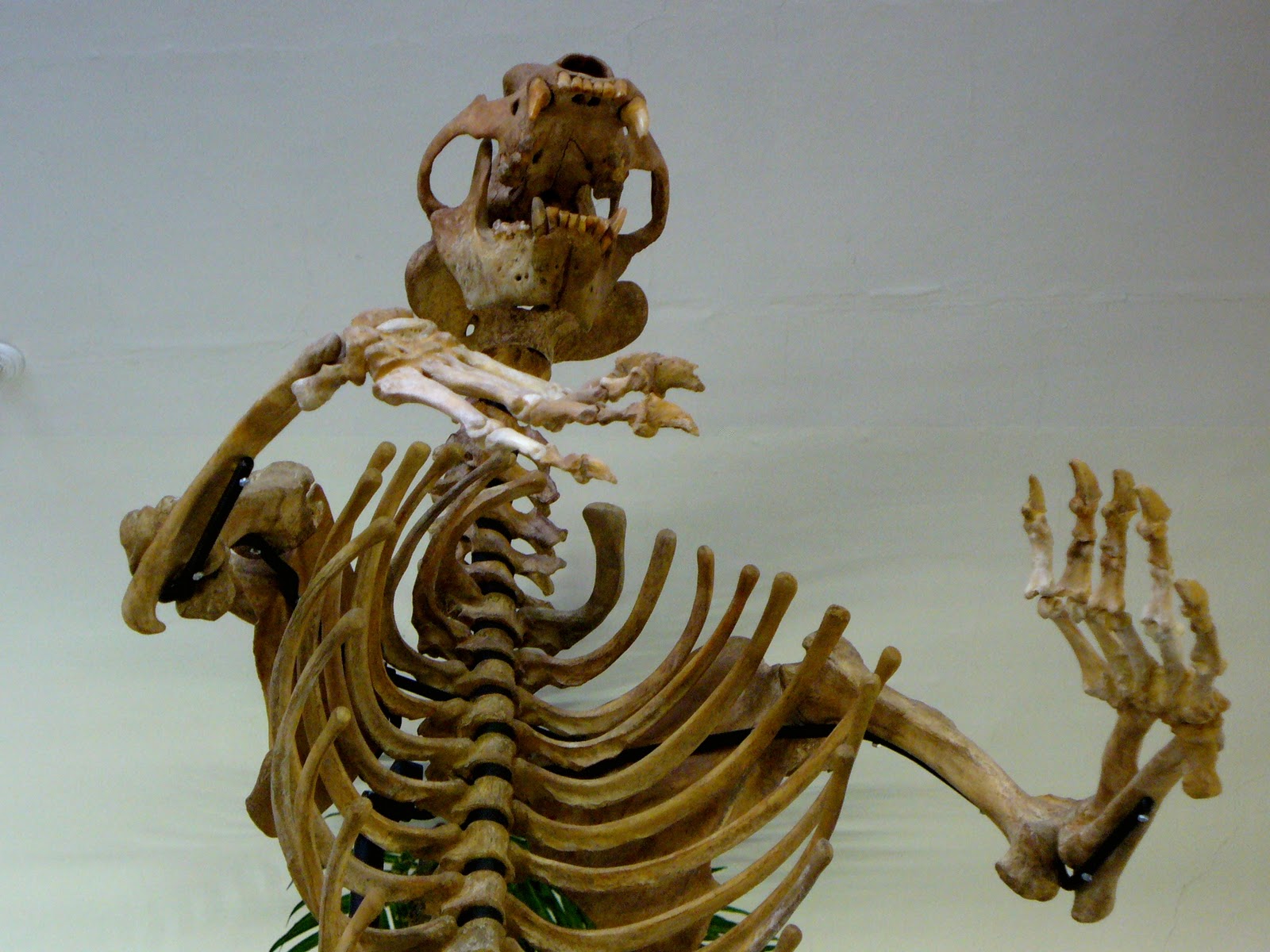
A Cave Bear skeleton in the typical (of museum displays) rearing posture.
Cave Bears lived in Pleistocene Europe (the Pleistocene epoch lasted from around 2.5 million to 11,700 years ago and covers the most recent series of repeated glaciations) and current evidence suggests they became extinct around 27,800 years ago. This means that they would have been encountered by humans and indeed they are depicted in cave art (albeit rarely e.g. at Les Combarelles cave in France). There is also possible evidence of Cave Bear worship by Neanderthals, such as at Drachenloch in Switzerland and Regourdou in France where the skulls of bears had clearly been arranged in and on man-made stone structures such as wall niches and a slab-covered pit. However, prehistoric anthropology, fascinating though it is, really isn't my area, so I'll stick with the more biological/ecological aspects. However, for an interesting overview of some aspects of human-Cave Bear interactions (focusing more on the earlier form of Cave Bear U. deningeri which disappeared around 100,000 years ago and may be an earlier species, a transitional subspecies or simply a pre-interglacial form of U. spelaeus), have a look at Stiner (1999). Taxonomic uncertainties aside, my interest was sparked when I bought a Cave Bear cheek tooth found in a cave in Romania, and dental evidence is where I will start.


Cave Bear cheek tooth, length 45mm.
This tooth is in pretty good condition (it's still shiny after about 40,000 years which shows just how tough tooth enamel is) and has an extensive grinding surface with a couple of large bluntly pointed cusps. Cave Bears lost their premolars as they evolved, a feature which has been used to suggest a highly herbivorous diet (e.g. Kurtén, 1976). The last premolar evolved as a molar (molarisation) which allowed tough plant material to be chewed more effectively (and hence more more food energy to be extracted) due to the increased number of cusps and cutting edges of the teeth, especially in the elongated last molar. Their teeth also show more wear than in most modern bears which again suggests a herbivorous diet with a large component of tough/fibrous materials, although detailed analysis indicates that tubers and other gritty foods were not a major part of their diet, unlike for modern Brown Bears U. arctos (Pinto Llona et al., 2005). However, varying threads of research in this area, including evidence for some cannibalistic scavenging (Pacher, 2000) has led to current scientific opinion tending towards Cave Bears being more herbivorous than modern bears of the genus Ursus, but still omnivorous to some extent. Recent re-examination of skull and tooth morphology (Figueirido et al., 2009) and analysis of the regional variation in bone isotope composition, especially nitrogen-15 (Richards et al., 2008; Trinkaus & Richards, 2008) both support this idea of omnivory and some variation in diet.

The same cheek tooth showing the pattern of the crown. The large grinding surface covers the left side of the tooth and the lower right side, with the pointed cusps to the upper right. The orange deposits in the grooves of the enamel are the remains of soil, although the right-hand end shows an area of worn (yellowish and not shiny) enamel at the base of the large cusp.
With advances in molecular biological techniques, the possibility of investigating cave bear genetics arose and in 2005, nuclear DNA extracted from a Cave Bear tooth around 42-44,000 years old was sequenced. This indicated that the Cave Bear was more closely related to the Brown Bear and Polar Bear U. maritimus than to the North American Black Bear U. americanus (Noonan et al., 2005) and supported earlier similar findings using mitochondrial DNA (Loreille et al., 2001). Interestingly, investigation of the fine structure of Cave Bear tooth enamel (the 'rods' or 'prisms' that form the basic units of enamel) shows that it retained carnivore-like characteristics despite the clear adaptation to a largely herbivorous diet. Thus, changes in broad dental anatomy driven by dietary specialisation can occur without the equivalent changes in enamel structure (von Koenigswald, 1992), meaning that Cave Bears had herbivore-shaped cheek teeth with carnivore-like enamel.
So, we have an extinct species of bear clearly adapted to a specialised herbivorous diet with some elements of omnivory and variation. As well as the genetic evidence mentioned above, its skeleton is similar to that of the modern Brown Bear, with the two species appearing to have diverged around 1.2 to 1.4 million years ago (Loreille et al., 2001) i.e. prior to the splitting of Brown and Polar Bears which may have occurred around 850,000 years ago, although this estimate is somewhat uncertain (Swenson, 2007). Males averaged 400–500 kg with females around half this weight at 225–250 kg (Christiansen, 1999), similar to the range for the largest modern bears, noting that they were larger during glaciations and smaller during interglacial periods (MacDonald, 1993), probably as an adaptation to adjust heat loss rate as larger animals have smaller surface area:volume ratios. The reason for its extinction is uncertain. It is unlikely to simply be due to its specialised diet and restricted geographical range ecologically 'marooning' it during post-glocial warming - after all, it had survived several similar changes in condition previously and there is possible genetic evidence of a decline starting some 25,000 years prior to its extinction (Stiller et al., 2010). Also, as noted above there is strong evidence for the species' ability to vary its diet. Instead, it is likely that there was a complex interplay of factors, possibly involving competition with humans for cave habitat, maybe specifically for hibernation sites as Cave Bears did not appear to use alternatives such as forest thickets and failure to find a hibernation site would lead to death. Despite numerous media reports taking the 2010 paper by Stiller et al. to be definitive evidence of competition with humans rather than changing climatic conditions to be the cause of Cave Bear extinction, there is still genuine scientific disagreement and research is ongoing. Further genetic work (Bon et al., 2011) does however show reduced genetic diversity from specimens in France originating from the period directly prior to extinction (genetic diversity is greater for specimens prior to this), again indicating a species under stress during human colonisation of the area - and the possibility of competition for hibernation caves.


References

Bon, C., Berthonaud, V., Fosse, P., Gély, B., Maksud, F., Vitalis, R., Philippe, M., van der Plicht, J. & Elalouf, J.-M. (2011). Low regional diversity of late cave bears mitochondrial DNA at the time of Chauvet Aurignacian paintings. Journal of Archaeological Science 38 (8): 1886-1895. 
Christiansen, P. (1999). What size were Arctodus simus and Ursus spelaeus (Carnivora: Ursidae)? Annales Zoologici Fennici 36: 93–102.
Figueirido, B., Palmqvist, P. & Pérez-Claros, J.A. (2009). Ecomorphological correlates of craniodental variation in bears and paleobiological implications for extinct taxa: an approach based on geometric morphometrics. Journal of Zoology 277 (1): 70–80.

Kurtén, B. (1976). The Cave Bear Story. Life and Death of a Vanished Animal. Columbia University Press, New York.
Loreille, O., Orlando, L., Patou-Mathis, M., Philippe, M., Taberlet, P. & Hänni, C. (2001). Ancient DNA analysis reveals divergence of the cave bear, Ursus spelaeus, and brown bear, Ursus arctos, lineages. Current Biology 11 (3): 200203.
MacDonald, D. (1993). The Velvet Claw: A Natural History of the Carnivores. BBC, London.

Noonan, J.P., Hofreiter, M., Smith, D., Priest, J.R., Rohland, N., Rabeder, G., Krause, J., Detter, J.C., Pääbo, S. & Rubin, E.M. (2005). Genomic Sequencing of Pleistocene Cave Bears. Science 309 (5734): 597599.
Pacher, M. (2000). Taphonomische Untersuchungen der Höhlenbären-Fundstellen in der Schwabenreith-Höhle bei Lunz am See (Niederösterreich). Beiträge zur Paläontologie 25: 11–85.
Pinto Llona, A.C., Andrews, P. & Etxeberrıa, P. (2005). Taphonomy and Palaeoecology of Cave Bears from the Quaternary of Cantabrian Spain. Fondacion de Asturias/Du Pont Iberica/The Natural History Museum, Grafinsa, Oviedo.
Richards, M.P, Pacher, M., Stiller, M., Quilès, J., Hofreiter, M., Constantin, S., Zilhão, J. & Trinkaus, E. (2008). Isotopic evidence for omnivory among European cave bears: Late Pleistocene Ursus spelaeus from the Peştera cu Oase, Romania. Proceedings of the National Academy of Sciences of the United States of America 105 (2): 600604.
Stiller, M., Baryshnikov, G., Bocherens, H., Grandal d'Anglade, A., Hilpert, B., Munzel, S.C., Pinhasi, R., Rabeder, G., Rosendahl, W., Trinkaus, E., Hofreiter, M. & Knapp, M. (2010). Withering Away 25,000 Years of Genetic Decline Preceded Cave Bear Extinction. Molecular Biology and Evolution 27 (5): 975978.


Stiner, M.C. (1999). Cave bear ecology and interactions with Pleistocene humans. Ursus 11: 4158.
Swenson, J.E. (2007). Økologi hos en voksende bjørnebestand – Forvaltning når bjørnen har kommet tilbake. Det Skandinaviske Bjørneprosjektet [in Swedish] [accessed 15/11/2011].

Trinkaus, E. & Richards, M. P. (2008). Reply to Grandal and Fernández: Hibernation can also cause high δ15N values in cave bears. Proceedings of the National Academy of Sciences of the United States of America 105 (11): E15.
von Koenigswald, W. (1992). Tooth enamel of the cave bear (Ursus spelaeus) and the relationship between diet and enamel structures. Annales Zoologici Fennici 28: 217227.

Wednesday, 9 March 2011

Entomology of Star Wars. Episode III: Bears in Space

Right, here it is - the third and final instalment of 'The Entomology of Star Wars' (unlike George Lucas I will not be tempted to produce a shiny but unsatisfying second trilogy). Following on from Episode II (exogorths - giant asteroid-dwelling worms), this time I decided to focus on the 'mynocks', the parasitic space-dwelling organisms that attach themselves to spacecraft to feed off their energy (they can feed on electrical, electromagnetic and stellar energy, and so may attach themselves to structures such as power cables or 'ion ports'), absorbing it through the suctorial mouth located between their eye stalks. They can also feed on the material of the hull, causing damage or even destruction; in turn they are eaten by exogorths and may survive for some time, flying around inside the worm's gut until finally digested.

A mynock attached to the Millennium Falcon, showing its leech-like sucker. Nice.
A little light reading ensures me that mynocks are silicon-based and fatally allergic to helium (they swell and die); also they lack any major organs and so reproduce by binary fission once they have fed sufficiently. Apparently, there have been other species (or subspecies), including one that had a normal mouth (rather than a sucker), and another that gave birth to live young. They do however have bat-like wings which they can use when in an atmosphere. So, what parallels can be drawn between the mynock and real-world organisms?

Firstly, as I've mentioned before, I'm not going to look at ideas surrounding non carbon-based life - there are plenty of bloggers looking at this already. Similarly, although mynocks can feed on ship hulls, I covered chemosynthesis and the mineral metabolism in Episode II. So, let's start with the most obvious aspect of mynock life (to me anyway) - their ability to survive in the vacuum of space. Although there are no known vacuum-dwelling real-world organisms, there is one experimental example which a lot of regular science readers may well be aware of - the tardigrade. For those who don't already know about this, here goes:

Tardigrades (Phylum Tardigrada) are microscopic to just-visible (adults range from 0.1 to 1.5mm) aquatic segmented animals with eight legs, sometimes known as 'water bears' or 'moss piglets'. They are well known for their ability to survive exceptionally hostile conditions, often more so than any other animal. Tardigrades are able to survive in extreme environments that would kill almost any other animal. Some have survived temperatures down to near absolute zero, up to approx 150°C and about a thousand times as much radiation as other animals, not to mention almost a decade without water. How do they manage this?

Tardigrade - a (tiny) bear in space!


Well, along with some other groups of organisms which can survive essentially complete dehydration, tardigrades accumulate large amounts of disaccharides ('double sugars' i.e. those with two molecular rings), especially sucrose and trehalose (Crowe et al. 1998). It appears that these sugars stabilise membranes and proteins when dehydrated, probably by hydrogen-bonding to polar residues in the dry macromolecular assemblages. This maintains dry proteins and membranes in a state similar to that seen when wet. It has been suggested that, as sucrose and trehalose form a glassy state when dry, glass formation (vitrification) is sufficient to stabilise dry biomaterials - however, Crowe et al. (1998) showed that both direct stabilisation and vitrification are required and that trehalose has properties linked to its crystal structure than may aid the stability and longevity of dehydration-tolerant organisms that contain it

Meanwhile, Jönsson et al. (2005) exposed tardigrades to high levels of gamma radiation. Without going into the results in detail, tardigrades do eventually die and/or become sterile, but the study concluded that their radiation tolerance may be due to currently unknown, but efficient, DNA repair mechanisms (unlike the  biochemical protection seen for their dessication tolerance). Then, in September 2007, tardigrades were taken into low Earth orbit on the FOTON-M3 mission and exposed to either the vacuum of space or both vacuum and cosmic/solar radiation. Despite these hostile conditions, where vacuum causes severe dehydration, while cosmic/solar radiation (with an unfiltered UV component) would be expected to cause considerable genetic damage. However, upon their return to Earth, many had survived and even laid eggs that hatched normally - they are the first animals to survive such exposure (Jönsson et al. 2008).So, they can protect their cells membranes and either protect and/or repair their DNA well enough to not only survive in space, but reproduce afterwards - our first parallel with mynocks, up to a point at least, but are there any others?

Well, I'm fairly sure there are no direct energy feeders (discounting photosynthesis of course!), unless you include the 'mystic woo' of psychic healing etc. Which I don't. However, the binary fission of a large organism is interesting - generally this is associated with small organisms such as Protozoa and Algae. However, it is also seen in some Myxomycetes ('slime moulds', but not Fungi). These are often soil micro-organisms, but in some species a single-celled, but multinucleate, 'plasmodium' is formed which is the final feeding stage, engulfing many kinds of small food items. Although essentially a single cell, these can be large - one example of Brefeldia maxima in North Wales can cover whole stumps, be a centimetre thick over a square metre and weigh up to around 20kg - possibly the largest cell known (Ing 1999), and its certainly feasible that such a structure could divide. So, we have our 2nd real-world parallel.

So, lastly on to the helium allergy... well, helium poisoning certainly can occur (e.g. if overused for 'squeaky voice' purposes), but although there's a general feeling of 'you can be allergic to anything', I've not been able to find anything about genuine helium allergies. It is pretty unreactive, and I suspect at least some of the anecdotal reports I found are actually allergies to the material the helium was held in (balloon rubber, latex glove powder etc). S, although a major anaphylactic event might parallel the effect of helium on mynocks, this remains an unproven 'maybe', and as Meat Loaf said, 'two out of three ain't bad'.

With that, I shall end my 'Entomology of Star Wars' trilogy, but fear not fellow invertebrate-SF nerds - I am starting to form ideas about B-movies and Iain M. Banks novels...

References

Crowe, J.H., Carpenter, J.F. & Crowe, L.M. (1998). The role of vitrification in anhydrobiosis. Annual Review of Physiology 60: 73-103.

Ing, B. (1999). The Myxomycetes of Britain and Ireland. Richmond, Slough.

Jönsson, K.I., Harms-Ringdahl, M. & Torudd, J. (2005). Radiation tolerance in the eutardigrade Richtersius coronifer. International Journal of Radiation Biology 81(9): 649-56.

Jönsson, K.I., Rabbow, E., Schill, R.O., Harms-Ringdahl, M. & Rettberg, P. (2008). Tardigrades survive exposure to space in low Earth orbit. Current Biology 18(17): R729-R731.

Monday, 21 February 2011

Woodpile wanderers and cohort-splitting

The value of dead wood as a habitat for invertebrates is well known (one of my 'static pages' covers this), so yesterday, while rummaging through our garden woodpile, I found what looked initially like a centipede about 20mm long, I felt compelled to put fingers to keyboard...

The first thing that leapt out was that it wasn't a centipede - it had two pairs of legs per segment (rather than one) and was therefore a millipede. The dorsal plates are splayed out sideways (the shape helps it push through the soil) and are coarsely sculptured along the back, and the legs and antennae are longer than in many other millipedes. Side on, it's clear why it's called 'flat-backed'.


Flat-backed Millipede


Side view showing the flat back

It's a common species, and is indeed known as the Common Flat-Backed Millipede (Polydesmus angustus). Found on roots and fruits (especially strawberries), it's often associated with all sorts of decaying plant material; dead leaves, compost, dead wood and so on. It's a native of north-west Europe, but has also been accidentally introduced to the south-east USA.

Although a common species, it does have some interesting life-cycle characteristics. Mating occurs from late spring into summer, then again from late summer to mid-autumn. Males usually mate only once, but females store the sperm from a single mating to produce several batches of eggs. Research by David (2009) showed that females born between May and August have a one-year life cycle while those born from late August onwards have a two-year life cycle (a strategy known as 'cohort-splitting', hence the title). A third type of life cycle ('interseasonal iteroparity' where iteroparity simply means reproducing more than once in their lifetime) was seen in a few females born late in the season.

Results from looking at the reproduction of individual females indicated that only annual females produced an appreciable proportion of biennial offspring from late August onwards; this means that life-cycle duration can't be genetically determined - cohort-splitting must therefore be driven non-genetically, supporting previous research by David et al. (2003) showing the effect of photoperiod (day length) on the life cycle of this species. As individual females reproduce for about two months, this automatically leads to a cyclical pattern of life-cycle duration (annual/biennial/annual) in the long-term progeny of any female.

Personally I find this fascinating, not least as an example of how a common species in an everyday habitat can surprise us with unuusal aspects of its biology or ecology. Wonderful! 

The telson and other posterior segments AKA 'the end'


Reference

David, J.-F. (2009). Female reproductive patterns in the millipede Polydesmus angustus (Diplopoda: Polydesmidae) and their significance for cohort-splitting. European Journal of Entomology 106(2): 211–216.

David, J.F., Geoffroy, J.J. & M. L. Célérier, M.L. (2003). First evidence for photoperiodic regulations of the life cycle in a millipede species, Polydesmus angustus (Diplopoda: Polydesmidae). Journal of Zoology (London) 260: 111–116.

Tuesday, 18 January 2011

Bushbuck: two species where there was one

Back in the day, the bushbuck was considered a single species, Tragelaphus scriptus, found in various habitats across much of sub-Saharan Africa. Recently however, genetic studies have indicated that T. scriptus is actually a complex of two distinct species, the Kéwel (T. scriptus) and the Imbabala (T. sylvaticus). This evidence shows that these two bushbuck species are more closely related to other tragelaphines than to each other; the Imbabala being closest to the Bongo (T. eurycerus) and Sitatunga (T. spekeii), and the Kéwel to the Nyala (T. angasii) (Moodley et al. 2009).

The Kéwel is found from West Africa, across the Sahel into East Africa, and as far south as Angola and the Democratic Republic of Congo (DRC). Meanwhile, the Imbabala is found from the Cape northwards to Angola, Zambia and East Africa, meaning that the two species’ ranges overlap in parts of Angola, DRC and East Africa.

The Kéwel is the smaller of the two, and shows clear stripes and patterning on a reddish to yellowish background; there is little or no sexual dimorphism in this ground colour. In contrast, the Imbabala shows considerable colour variation with geography and habitat, especially in males (yellow to red-brown, through brown and olive to almost black), and only the most genetically ancient of populations (from Angola, Zambia, southern DRC, Botswana and northern Zimbabwe) have any significant striping. Even in these cases the horizontal stripe, where it exists, is formed of a series of spots rather than the solid striping of the Kéwel. never occurs. Mountain-dwelling forms of the Imbabala (Gregory Rift Highlands, Mt. Elgon, Imatong Mountains and Ethiopian Highlands) appear larger and are dark with little or no pattern. Until recently, most bushbuck studies focused on the Imbabala, hence little was known about the biology of the Kéwel beyond what could be obtained from museum specimens and hunting trophies.

Imbalala bushbuck from Zimbabwe (courtesy of Graeme Guy). For a kewel image from The Gambia see here.
Both species are primarily browsers, but will eat other plant matter too. They can be active at any time of day, although are more likely to be nocturnal near humans; their most active times are however early morning and parts of the night, so may appear nocturnal in any case. Most are solitary, with some living in pairs; all have a ‘home range’ of around 5 hectares in the savannah (larger in forests), although these ranges do overlap.

Although the split into two species is fairly well understood (even if most non-scientific sources still refer to a single ‘bushbuck’), the more detailed taxonomy remains disputed with numerous potential subspecies and ecotypes having been described. For example, analysis of mt-DNA sequences (cytochrome b and control region) by Moodley & Bruford (2007) identified 23 phylogenetically distinct groups (‘ecotypes’) whose distribution correlated well with the pan-African eco-regions described by Olsen et al. (2001). 19 of these ecotypes corresponded with previously suggested subspecies, while six other haplotypes were newly recognized forms in the Volta region, Niger, Angola. and Luangwa and Zambesi Valleys. However, further research is onging to clarify the taxonomic status of bushbuck species, subspecies and ecotypes, so the situation is likely to remain somewhat fluid for a while – however, this does provide an opportunity to link the use of genetics in taxonomy to large-scale conservation in Africa, given the widespread distribution of bushbuck (in the broad sense) and apparent more local/region distribution of subspecies and ecotypes (Wronski 2009).

References

Moodley, Y. & Bruford, M.W. (2007). Molecular Biogeography: Towards an Integrated Framework for Conserving Pan-African Biodiversity. PLoS ONE 2(5): e454. doi:10.1371/journal.pone.0000454

Moodley, Y., Bruford, M., Bleidorn, C., Wronski, T., Apio, A., & Plath, M. (2009). Analysis of mitochondrial DNA data reveals non-monophyly in the bushbuck (Tragelaphus scriptus) complex Mammalian Biology - Zeitschrift fur Saugetierkunde, 74 (5), 418-422 DOI: 10.1016/j.mambio.2008.05.003

Olson, D.M., Dinerstein, E., Wikramanayake, E.D., Burgess, N.D. & Powell, G.V.N. (2001). Terrestrial ecoregions of the world: a new map of life on earth. BioScience 51: 933-937.

Wronski, T. (2009). Bushbuck, harnessed antelope or both? Gnusletter 28(1): 17-19.

Thursday, 6 January 2011

Britain's jawless wonders: brook lamprey ecology and taxonomic status.

Lampreys (family Petromyzonidae, ‘stone-suckers’) belong to the Agnatha, (‘jawless’), the most primitive of all living vertebrates. They have no lower jaws and the mouth is surrounded by a round sucker bearing circles of rasping teeth. They are eel-like in shape but have neither paired fins nor scales and their skeletons are entirely cartilaginous. There are three species of lamprey in Britain: brook (Lampetra planeri), river (Lampetra fluviatilis) and sea (Petromyzon marinus), but having seen L. planeri nest-building in a nearby river (the R. Itchen in Hampshire), it is this species that I want to focus on here.


Brook lamprey (Lampetra planeri) nest-building in the River Itchen, Hampshire, UK.

L. planeri  is the smallest of the three British species at 13–15 cm long. The teeth are blunt and less developed than in the other more predacious species, and it does not feed as an adult. Larvae (‘ammocoetes’), are semi-translucent and dull grey-brown in colour, though a 'golden' form does exist with reduced pigmentation. Larvae occur in suitable silt beds, mainly in running water but sometimes in large numbers in silt banks in lakes. Although not common, it is the most abundant and widespread British lamprey and its distribution can be seen in the maps below for the National Biodiversity Network (NBN); the larger-scale map shows Hampshire and surrounding areas. However, due to a decline in several parts of Europe, it has some legal protection including being long-listed in the UK Biodiversity Action Plan.


British distribution of L. planeri (from NBN as of 6th Jan 2011)

Distribution of L. planeri in and around Hampshire (from NBN as of 6th Jan 2011)

Usually cryptic and nocturnal, L. planeri (like most lampreys) are rarely seen except at spawning time (April & May) when they move into shallow, clear water during daylight to start their complex, communal nest-building activities - the photo above was taken in April 2009. As spawning approaches, adults move from silts and migrate upstream at night, often in large numbers, until they reach suitable spawning grounds. These are areas of small stones and gravel in flowing water where the current is present but not too strong. When the water reaches 10–11ºC they tend to spawn at the lower ends of pools, just where the water is starting to break into a riffle. The nest may be constructed by a dozen or more adults moving stones with their suckers and is normally an oval depression 20–40 cm across and 2–10 cm deep. Females produce about 1,500 eggs each and hatched larvae (3–5 mm long and blind) drift downstream to burrow in suitable areas of silty sand. The adults die soon after spawning. Larvae live for around 6½ years in Britain, filtering fine organic particles from the silt. Metamorphosis occurs during July to September, after which they are more silvery, though the back remains dark. They also develop teeth and full vision, though adults do not feed.

Upstream migration can only occur in the absence of barriers, either natural (e.g. waterfalls) or man-made (e.g. dams, weirs or polluted areas). Little is known about their requirements in terms of water quality and quantity, though some limited pollution appears to be tolerated if it does not lead to substrates being smothered. They need suitable conditions (e.g. substrate type) in spawning areas and nursery habitat, and rivers should not be changed to produce excessive cover or fast flow. Channelisation and some aspects of management for angling (e.g. dredging of pools and construction of weirs) has been damaging to lampreys, mainly through habitat destruction, with potential impacts from use as bait. The removal of riffles and associated spawning gravels, and the dredging of silt beds can entirely eliminate lampreys from a river. Similarly, water abstraction and land drainage can produce unstable habitats with variable water levels which flood and disturb spawning gravels and nursery silts at some times and dry out at others. Climate change is likely to produce similar problems, with heavy rain in the autumn and winter, and drought in the summer.

Also, although L. planeri and L. fluviatilis are generally treated as separate species, there is some genetic evidence that this might not be the case. Schreiber and Engelhorn (1998) found very little difference in the DNA content between both species suggesting L. fluviatilis may just be an anadromous form of the relatively 'stationary' L. planeri, with gene flow inferred between them. Their taxonomic relationship and status is still a matter for debate although further research continues to suggest that they may not be truly separate species. For example, Lesne et al. (2010) show a high incidence of communal spawning of the two species in a French river, suggesting that they are less reproductively isolated than previously believed.


From a practical conservation perspective, L. planeri (whether or not it is a species separate from L. fluviatilis) does have some key requirements, but nothing that a little more considerate and thoughtful river/habitat management shouldn't be able to provide in abundance, and which would in turn help mitigate the likely effects of climate change.

References

Lasne, E., Sabatié, M., & Evanno, G. (2010). Communal spawning of brook and river lampreys (Lampetra planeri and L. fluviatilis) is common in the Oir River (France) Ecology of Freshwater Fish, 19 (3), 323-325 DOI: 10.1111/j.1600-0633.2010.00428.x

Schreiber, A., & Engelhorn, R. (1998). Population genetics of a cyclostome species pair, river lamprey (Lampetra fluviatilis L.) and brook lamprey (Lampetra planeri Bloch) Journal of Zoological Systematics and Evolutionary Research, 36 (1-2), 85-99 : 10.1111/j.1439-0469.1998.tb00781.x

Further reading


  • More on lamprey ecology can be found here.
  • An identification guide to British lampreys can be found here.
  • A guide to monitoring lamprey can be found here.