Welcome

Welcome to my blog

This is where I post various musings about wildlife and ecology, observations of interesting species (often invertebrates)
and bits of research that grab my attention. As well as blogging, I undertake professional ecological & wildlife surveys
covering invertebrates, plants, birds, reptiles, amphibians and some mammals, plus habitat assessment and management
advice
. I don't work on planning applications/for developers. The pages on the right will tell you more about my work,
main interests and key projects, and you can follow my academic work here.

Tuesday, 31 January 2012

What's in the box? No.13 - new to Nottinghamshire

The most recent coleopterous arrival by post came beautifully carded inside the lid of a screw-top plastic pot complete with a label detailing the location, collection date and name of the person recording it. It came from a reserve warden who had been in touch with me because they had identified the beetle as Longitarsus dorsalis, a species that did not appear to have been recorded before in the county in which they found it (Nottinghamshire) and wanted the identification confirmed (or otherwise).

The neatly set specimen of Longitarsus dorsalis as it arrived by post - carded inside the lid of a screw-top plastic pot.
Now onto identification. The beetle is about 2.5mm long (as ever, excluding appendages), with dark elytra bearing broad orangey side-stripes, a similarly orange pronotum and a dark head and appendages.

Dorsal view of L. dorsalis
Within the genus Longitarsus, this coloration is only known from L. dorsalis in Britain and the genus is easy to determine as the first tarsal segment is more than half the length of the tibia. So, it seems that the identification is straightforward to confirm, but it is worth looking more closely to see some other features associated with the species (some more details and further images can be found here).

Hind leg of L. dorsalis showing key features
In this photo, the long first tarsal segment is clear to see (red line) and the tibial spur (green line) is shorter than the maximum thickness of the tibia (sometimes this can be difficult to tell as the spur length may be very close to the max tibial width). The upper surface of the tibia is flat (too dark to tell here, but it is) and the outer edge of the tibia has a fringe of short flat bristles (red arrow).

Pronotum of L. dorsalis
The pronotum is orange, usually with at least a dark patch towards the front (clearly seen above). The pronotum also has a fine but distinct rim along the rear edge (also visible above) and sides. The pronotal punctures are fine and between them there is finer microsculpture. The elytral punctures are coarser than those on the pronotum and the elytra themselves have definite 'shoulders'.

Head of L. dorsalis
The head is also densely microsculptured and although this isn't visible in the photo above, you can see the coarse punctures along the edge of the eye.

Side view of L. dorsalis showing the epipleura narrowing towards the rear (red lines).
In side view, the epipleura (lower edges of the elytra) narrow towards the rear, especially behind the mid-point (approximately where the left-hand red 'I' is in the photo above). Lastly, I want to look at the aedeagus (below) which is often a useful diagnostic tool when identifying some trickier-to-separate beetles. Here, the tip unfortunately broke off during dissection (though it did have a small blunt point), but the sides clearly curve inwards and the shape matches the better specimen illustrated here. The parameres (lateral lobes) can be seen as the dark V-shaped structure near the bottom. Interestingly, the curved sides of the aedeagus, an important feature, are not clearly shown in the usually excellent (and expensive) work by Warchałowski (2003).

Aedeagus of L. dorsalis

This clearly indicates that the specimen is of L. dorsalis which is important as it is the first time it has been found in Nottinghamshire, and is nationally scarce in the UK (Notable B). It is mainly found on calcareous or sandy soils, is associated with ragworts (Senecio), and has become scarce due to the loss of its habitats (conversion of grassland to agriculture, infilling of quarries, habitat succession such as in once-open woodland rides and clearings, grassland 'improvement' through fertiliser application, herbicide use and woodland clearance). Open conditions with ragwort are required and conservation measures can be straightforward e.g. retaining some open areas in woodland through rotational cutting, or grazing is some other situations (Hyman, 1992). So, it's good to see this species in a previously unrecorded location, especially as several individuals were seen with just one sent to me for identification.

References

Hyman, P.S. (1992). A Review of the Scarce and Threatened Coleoptera of Great Britain. Part 1. JNCC, Peterborough.
Warchałowski, A. (2003). The Leaf-beetles (Chrysomelidae) of Europe and the Mediterranean Area. Natura Optima Dux Foundation, Warsaw.

Monday, 30 January 2012

Observations of Macleay's Spectres IV: moulting and leg regrowth

Parts I-III of this series have looked at different life-cycle stages and sexes of the Macleay's Spectre stick insect Extatosoma tiaratum. In this 4th part however, I want to look at processes and structures relating to ecdysis (moulting) and the regrowth of lost legs.

In invertebrates such as stick insects there is an incomplete metamorphosis with a series of nymphs (instars) that gradually increase in size but are approximately the same overall form as the adult. During each instar, the next larger 'skin' is grown wrinkled inside the existing one - when ready, hormonal effects detach the skin layers from each other and the old one splits along predefined lines of weakness. The next instar can then emerge, leaving an empty skin or 'exuvium'. This contrasts with groups that show complete metamorphosis where the larva develops into a pupa before emerging as an adult (imago) with a very different appearance - the most well known example of this is probably the butterflies.

In E. tiaratum, the soon-to-moult nymph clings to foliage or twigs with its legs, and the old skin splits lengthways along the dorsal midline of the thorax starting at the back of the head. The insect then pulls its front half free and leans backwards through this split, hanging by its still attached abdomen - the whole process takes from 30 minutes to two hours (Brock, 2000).

Final instar female E. tiaratum moulting.
The abdomen is slowly freed using muscular contractions to pull away from the old skin. When nearly free, the insect grips a suitable object and hangs abdomen-down as the new skin hardens and the insect takes in air to expand itself and stretch out the previously wrinkled skin - thus its overall size increases. This arrangement means that in captivity a container needs to be at least three times as high as the length of the extended insect; if the height is insufficient, the soft abdomen may press onto the floor of the container and harden with an unwanted bend which presumably may cause problems with digestion/waste elimination and reproduction if too pronounced. Apparently it is possible to straighten out unwanted bends by using a glued-on matchstick as a splint (Brock, 2000), but I have no wish to try this!

The picture above shows that the rear left leg is very small - this is due to a leg being lost during a previous moult. This highlights the risks associated with moulting and in this case was due to the old and new skins not separating around this leg. Having watched the otherwise fully moulted insect attempting to remove the still-attched skin for some time, I was forced to intervene and the leg was removed. However the loss of one (or even two or three) legs does not unduly affect the insect (in the wild it is a useful way to escape predators who are left with just a single twitching leg). Lost legs do regrow to some extent with successive moults but as seen here are smaller, starting as a thin black stump after one moult. Interestingly, research on another stick insect species (Sipyloidea sipylus) indicates that regrowing legs reduces adult wing size and hence flight performance, showing that there is a trade-off between development of legs and wings due to limited total resources for appendage growth and that this may have affected the evolution of this group (Maginnis, 2006) which has lost and regained wings on a number of occasions throughout its evolutionary history.

Usually, the old skin is eaten as the first post-moult meal, but occasionally it is lost (e.g. if it falls from its point of attachment) and subsequently ignored. I have also seen larger specimens eating the shed skin of another recently moulted individual. However, when a skin remains uneaten it is a useful way of investigating the finer structure of these insects and the moulting process.

A whole moulted skin of a male E. tiaratum showing the long wing-buds. This is approximately 40mm long although the curled abdomen means the insect was around 60mm long prior to moulting.
One feature visible here and in the top photo is the series of thin white threads around the head. These are not antennae (which are brown and can be seen underneath the head); instead they are the air tubes that form the trachael or breathing system of the insect and run internally from small air holes or spiracles along the sides of the abdomen. These are attached to the abdomen at the spiracles and so when the insect moults these also have to be pulled free and thus are found inside-out and external rather than internal.

The dorsal split through which the insect emerged during moulting. The air tubes are clearly visible here including their points of attachment at the spiracles.
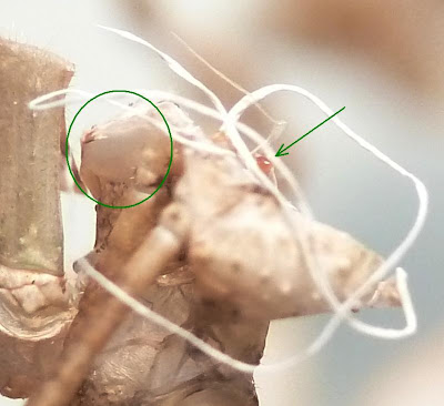
Lastly, it is important to remember that in insects the eyes (both compound and simple/ocelli) and structures such as mouthparts and antennae are covered by part of the exoskeleton and so are also involved in moulting. The photo below shows the moulted surfaces of both types of eye - the arrow indicates and ocellus while the circle surrounds a compound eye.

The moulted head of E. tiaratum showing the surfaces of both types of eye.


References

Brock, P.D. (2000). A Complete Guide to Breeding Stick and Leaf Insects. Kingdom, Havant.
Maginnis, T.L. (2006). Leg regeneration stunts wing growth and hinders flight performance in a stick insect (Sipyloidea sipylus). Proceedings. Biological sciences / The Royal Society 273(1595): 1811-1814.

Thursday, 26 January 2012

Observations of Macleay's Spectres III: the girls

Following sections covering eggs and early nymphal stages and mature males, the third part of this series on Extatosoma tiaratum, the Macleay's Spectre, looks at mature females.

Female E. tiaratum with front legs raised.
As late-instar nymphs and adults, females are structurally very different from males (sexual dimorphism). They are larger - up to 150mm long with males around 100mm, broader and much heavier - up to around 30g (a lot for an insect and females often drag the spiny underside of their abdomen along the surface they are walking on, sometimes producing a clear scraping noise as they move). Females also have many more spines and flanges, including a more pronounced 'head-dress' of short blunt spines. In fact, in E. tiaratum, the differences between males and females are so pronounced that they were originally described as separate species (Hadlington & Johnston, 1998)! Details of female genitalia are given by Heather (1965) and note that from a sample of seven specimens there were on average 151 ovarioles (tubes forming the paired ovaries) with an average of seven oocytes (immature egg cells) each. The bursa copulatrix (the sac-like organ where the spermatophore or 'sperm packet' is stored after copulation) bears around 20 blind tubules and there appears to be no distinct spermatheca (sac for sperm storage) or other sclerotised (hardened) part of the genitalia. This matches the observation (see Part II) that sperm transfer is by spermatophore.

Flanges and spines on the leg of a female.
The spine-covered abdomen of a large female.
Side view of a female's head showing the oval extension of the top with its 'head-dress' of short spines. Also note that the camouflage includes marking breaking up the shape of the eye.
The head of a large female showing small wart-like bumps as well as a clear view of the mouthparts.
Around the same time my males changed to a brick-red colour, my two large females developed a pinkish-lilac tinge as shown in the photo of the abdomen above. This may be an indication of sexual maturity or possibly gravidity (containing eggs). Also, following their final moult, the adult females became noticeably more aggressive, making for interesting cage-cleaning sessions... This aggression included general attempts at evading capture (fleeing or dropping out of reach; previously these had not occurred often) plus active attempts to use spines as weaponry (such as pinching with the inner edge of a bent leg, nut-cracker style), including against unwary males that strayed too close soon after an angry female was replaced in the cage. I have seen one male with what appears to be a small healing puncture wound (i.e. a hardened drop of what I imagine to be dark haemolymph) on the ventral side of the thorax though whether this was caused by female aggression or an accident with a bramble thorn I do not know.

A female nymph already showing spines and flanges.
Side view of a late-instar female nymph.
As this pair of photos shows, the female structure develops at a relatively early stage, essentially just getting larger with each moult. This contrasts to some extent with males which show a number of significant structural changes following their final moult.

A big girl perching on my wrist. She is mighty!
So, I started this series of posts with eggs, and can return to this topic as two days ago one of the large females began laying eggs. This is not a precise process in E. tiaratum as eggs are flicked up to a few feet (maybe a metre or so) by twictching the abdomen when the egg is laid (this also occurs with frass AKA insect faeces which takes the form of dry 3mm x 5mm oval pellets of plant material - in males, frass is long and thin, approx 1mm x 5mm). Fortunately as my insects are in cages, the eggs are easy to collect so I hope to have another generation relatively soon.

Tip of a mature female's abdomen showing an egg about to be laid. Note the dorsal spines on the abdomen - they are splayed outwards which may be an adaptation to accommodate a male during mating.

A female nymph doing her favourite thing - eating bramble leaves, nom nom nom.

It's busy during cage-cleaning time.

References

Hadlington, P & Johnston, J.A. (1998). An Introduction to Australian Insects (revised ed.). University of New South Wales, Sydney.
Heather, N.W. (1965). Studies on female genitalia of Queensland Phasmida. Australian Journal of Entomology 4(1): 33-38.

Tuesday, 24 January 2012

Observations of Macleay's Spectres II: the boys

Having looked at the early stages (eggs and small nymphs) of my Macleay's Spectre stick insects (Extatosoma tiaratum), it's time to move on to looking at the older males. As they develop through a series of nymphal stages (instars), moulting and expanding in order to grow, they develop small spines and flanges, but most obviously a pair of wing buds. In their final (5th) instar, these are quite obvious and when they moult, emerging as an adult, these develop into full pleated wings with long 'coat-tail' covers. 

An adult male showing its long wing covers.
An adult male opening its wings just as it is about to take flight.
Once they have dried their wings (much like butterflies and other winged insects do), the males are able to fly strongly, and in the wild do so in order to find food and females, or to evade predation. However, I found that initially they were not very active apart from showing increased aggression when handled. During their final moult they also develop longer, curved antennae and larger, more protruding eyes, presumably used to find females both by scent at a distance and then visually when nearer. The wings fold along radial pleats (a bit like a parasol) and have a spotty pattern, and the neck is long and flexible at the joint with the thorax.

Head-on shot of a male in a threat posture showing well developed eyes and antennae.
The males remain well camouflaged, moving in a similar manner to leaves in the wind. For a few weeks they retained their variable colouring - some males were greenish, others brown or greyish. However, after this time a change seemed to take place with most darkening to a reddish-brown colour, and becoming more willing to fly - most evenings, the vivaria are opened and some males readily walk onto my outstreched hand and then launch (this is preceded by a subtle but definitie raising of the body into a launch posture), often having climbed onto my head or shoulder first. Around the same time, the first males could be found mating with the large adult females (more about them in Part III). So, it appears - albeit anecdotally - that this change in colour signals sexual maturity. If so, this is interesting as it does not seem to be associated with increased aggression either towards me (if anything they seem less aggressive when handled and simply fly more readily, though they may simply have habituated to handling, and some definitely avoid handling if possible) or other males. A number of males can be seen clustered around a female on occasions, but I have witnessed to overt aggression, though I have to assume that there is some form of competition to mate - maybe I need to observe what they are doing at 3am...

A male showing a dark red-brown colour and the long neck. The bright dot on top of the head is one of the male's ocelli (simple eyes).
A yellow-brown late (5th?) instar male nymph with the long wing-buds just visible behind the right middle leg.
Another late-instar (again, 5th?) male nymph, this one pale green in colour, again with the wing-buds visible.
When mating (or preparing to do so), males lie lengthwise along the back of a female (in the usual legs-outstretched 'stick' position) and both have genitalia at the rear of the flexible abdomens which bend to fit. The function of the long male neck with flexible articulation then becomes evident; females often arch backwards when feeding or moving and this forces the male's head backwards - the flexible neck allows him to remain in position without damage.

Males using my wife as a climbing-frame/launchpad while their cage is being cleaned. Just prior to this photo being taken, one of the males appeared to be trying to mate with her hair-grip (it has strong legs and a handy, accommodating central groove...)

Sperm transfer takes place in the form of a spermatophore - a packet of sperm in a hard 'shell' which novice insect keepers sometimes mistake for eggs. The spermatophore of E. tiaratum was noted by Clark (1975) and has been well documented since, but it was not until relatively recently that review of research and observations (e.g. Bragg, 1991) concluded that this structure provided the usual method of sperm transfer in the order Phasmida (AKA Phasmatodea). I recently collected an E. tiaratum spermatophore from the floor of one of my containers at home. The photo below shows the outer structure - the thread attaches it to the male during transfer to the female and the sperm-containing sac is 2.5-3mm in diameter, the whole being white with a pink tinge especially where the thread attaches to the sac.

Spermatophore of E. tiaratum
That's all for the males, but why not check out Part III: the girls...

References

Bragg, P.E. (1991). Spermatophores in Phasmida. Entomologist 110(2): 76‑80.
Clark, J.T. (1975). A conspicuous spermatophore in the phasmid Extatosoma tiaratum Macleay. Entomologist's Monthly Magazine 110: 81-82

Sunday, 22 January 2012

Observations of Macleay's Spectres I: early stages

Last summer, I was given an unusual birthday present - a 35mm film canister full of eggs of the Macleay's Spectre stick-insect Extatosoma tiaratum, sometimes known as the Giant Prickly stick-insect. These are found mainly in the Australian forests of Queensland and New South Wales where they feed on the foliage of eucalyptus trees, although in captivity they eat a range of unrelated species - one popular food is bramble (Rubus fruticosus agg.) which is what I use as it is readily available for free. However, young bramble leaves can contain noxious chemicals that make them unpalatable or even toxic, especially to young insects - I tend to remove these, and when some have been included by accident with more palatable older leaves, the insects ignored them.There is plenty of information about this species (e.g. from the Phasmid Study Group) as they are popular pets for those who like their companion animals to be six-legged, but I have made some observations which may be of interest. So, let's start at the beginning - with eggs...

Egg of E. tiaratum with the lid (operculum) open.
Close-up of the operculum showing the translucent sealing membrane around the edge. There is another membrane below this which seals the opening to the egg and which is broken to allow emergence by the first-instar nymph.
The eggs are a few mm long, oval with a sculptured ridge along one long axis, and a round lid (operculum). They are speckled with various shades of yellow and brown, camouflaging them to look like seeds when they fall to the forest floor in the wild. In those I have at home, the eggs are a pale yellow-brown when freshly laid and dry, but when placed on damp tissue to (eventually) hatchm they become a dark brown - presumably this aids camouflage with dry soil conditions also being paler and darkening when there is rain. When females lay eggs, they often flick them several feet by twitching their abdomens, presumably to aid dispersal  - the function of this might be to prevent a cluster all being eaten in one go by a predator. The eggs have a coating (made of lipids and other organic materials) which is edible to ants and which induces them to take the eggs to their colony and eat this coating. The eggs remain otherwise intact and the ants dump them on the colony's waste pile where they hatch. Thus the eggs are likely to become clustered following the initial dispersal during laying but will have spent much of their time in an ant colony where the chance of predation is much reduced. Luckily, the eggs can hatch without the attention of ants which means that those missed by foraging ants and of course those in captivity remain viable.

Unsurprisingly, given their birthplace, E. tiaratum hatchlings (forst-instar nymphs) are ant-mimics, specifically of the large, long-legged 'spider ants' in the genus Leptomyrmex.

Recently hatched nymph showing the orange head and black body providing its mimicry of Leptomyrmex ants.
The orange head and black body, as well as overall shape closely mimics that of ant species such as L. darlingtoni, L. erythrocephalus and L. rufipes and so benefit from appearing similar to these toxic species (the sting has degenerated in this group of ants and they instead secrete a chemical repellant).

When my specimens hatched, I couldn't help but notice that the nymphs were highly active (a well-known characteristic) and that their preferred direction was definitely up. This makes sense as they hatch on the ground but are foliage feeders and so need to climb quickly into trees to find food. After their first moult, they lose their ant-mimic coloration and develop one of a range of camouflage colours - oranges, yellows, browns, greys and greens - and they also become much less active, more like the typical behaviour of a stick-insect. They do make swaying movements to mimic the motion of leaves, especially when disturbed, but otherwise spend much of their time stationary or moving slowly as they feed, though being nocturnal much of their activity goes unobserved (or would if I wasn't so nosy). The image below shows three nymphs at different stages and with different colours. In subsequent parts of this series, I'll be looking at changes in colour and behaviour (in males and females, highlighting sexual dimorphism) plus the process of moulting.

Three nymphs showing different colours and stages of development. The two smaller ones are 2nd instar nymphs, the larger one a 3rd instar.

Monday, 16 January 2012

Teeny-tiny toadstools - or are they?

Yesterday I was pointed towards a log cut from a fallen tree in my in-laws' garden. On it was a fairly dried-out growth of moss and on the leaves, what looked like some tiny (1mm) fungal caps. Now, I'm no mycologist (or bryologist), but I do find microfungi intriguing and happen to have a copy of Ellis & Ellis (1998) which is the standard (only?) work covering British microfungi on substrates other than vascular plants - for those you need Ellis & Ellis (1997).

The specimen - the caps are about 1mm in diameter.
A slightly closer view showing the wrinkled stalks.
The moss is fairly dry and a bit of a miserable specimen without capsules, but looking at the excellent Atherton et al. (2010), I think that it is the common and widespread species Brachythecium rutabulum (sometimes known as the Rough-stalked Feather-moss). The fungus itself is white with a split cup-like cap and a wrinkled stem that widens towards the base. The cap is granular beneath and bears hairs in the upper/outer surface.
Two of the fruiting bodies showing details of stems and caps.
Side view of a fruiting body showing the split cap and granular spore-bearing structures.
Some moss-epiphytic microfungi have clear fringes of hairs around the edge of the cap. This doesn't appear to be the case here - instead there appears to be an irregular tufting of hairs on the top of the cap with a few at the edge (I wondered if they were the hyphae of another even smaller fungus, but I don't think so).

Side view of the cap showing tufts of hairs.

High-power image of the cap showing branching hairs.

Lastly, spores are an important feature used in the identification of microfungi. I collected some of these on a slide and they are clearly almost spherical and reddish-brown in colour, and each is about 10um in diameter.

Spores (x100 magnification)

So, the overall size, colour, spore colour/shape/size, habitat, hairs and other features lead me to tentatively identify this as Chromocyphella muscicola, a species with caps up to about 3mm diameter which is associated with mosses on bark. However, having consulted with a mycologist from the Hampshire Fungus Recording Group, it appears that C. muscicola doesn't produce stalks like this. Other superficially similar genera such as Leptoglossum also differ from this specimen in one or more ways e.g. spore shape, cap hairs. And so, it was suggested that this might not be a true fungus at all, but instead a myxomycete or 'slime mould' where the globular sporangium had broken open to reveal the network of spore-bearing hairs known as the capillitium.

I have posted about myxomycetes before (e.g. here and here) but it still a group of organisms I know little about, fascinating as they are - taxonomically protozoans but traditionally treated as 'honorary' Fungi. So, swapping books to look at Ing (1999), it soon became clear that this was indeed a 'myxo'. It also happened to be one that was relatively easy to identify from the keys and descriptions - the pale colour, size, stalk, open split 'cap' with the hairs of the capillitium, and spores (shape, size, colour, texture). This process also taught me that an important identification feature can be the shapes, colours and sizes of nodes of the capillitium i.e. structures about the size of a spore where hairs meet and branch.

These features all combined to indicate that this is the myxomycete Physarum nutans - a common and widespread species, though rarely found on mosses (it is usually on dead wood or the bark of live trees). It is very similar in appearance to Didymium squamulosum but they can be separated by features such as the spores (in D. squamulosum they are dark brown in transmitted light whereas in P. nutans, as here, they are pale brown). So, an interesting process for me, looking at an unfamiliar group - the only way to learn more about species identification!

References

Atherton, I., Bosanquet, S. & Lawley, M. (eds.) (2010). Mosses and Liverworts of Britain and Ireland: A Field Guide. British Bryological Society.
Ellis, M.B. & Ellis, J.P. (1997). Microfungi on Land Plants: An Identification Handbook. Richmond, Slough.
Ellis, M.B. & Ellis, J.P. (1998). Microfungi on Miscellaneous Substrates: An Identification Handbook. Richmond, Slough.
Ing, B. (1999). The Myxomycetes of Britain and Ireland: An Identification Handbook. Richmond, Slough.

Friday, 13 January 2012

What's in the box? No.12 - when typical species ain't so typical

Back to the small beetles today - I've just finished identifying all the specimens that I received by post a while back (yay!) - most were fairly common, though a few such as the chrysomelid Phyllotreta punctulata were scarcer (in Britain at least). However, 'common' does not necessarily equal 'easy to identify' and here are a couple of examples from the family Chrysomelidae (leaf-beetles). The first is a deep metallic blue beetle about 4mm in length (without appendages).
The first specimen, a shiny blue chrysomelid beetle about 4mm long.
One very clear feature here is the pronotum being considerably narrower than the elytra. The length of the pronotum is about 0.4 that of the elytra and the elytra are about 1.6-1.7 times as long as they are wide. The overall form is distinctive and indicates it is a member of the subfamily Criocerinae.

A close-up of the pronotum with key features indicated.
 Looking more closely, the green arrow on the left points to a rounded but definite angle (i.e. the side if the prronotum is not evenly rounded) while the red bracket shows that the constriction near the rear of the pronotum lines up with the furrow across its upper surface. These features separate it from the genus Lema (which would have the constriction near the rear of the pronotum and not in line with the furrow, and would also have sharper angles on the side), and hence show it to be in the genus Oulema - there is a good page separating L. cyanella and O. obscura here which also covers some of their taxonomic confusion. In Britain there are three Oulema species which are this colour - obscura, septentrionis and erichsoni.

O. obscura (often known as O. gallaeciana in continental Europe) is generally considered to be a fairly short species with a pronotum 0.3 times as long as the elytra and with elytra 1.25 times as long as wide. So, using these features, it would appear to be one of the other two, more elongate, species. However, there is a problem here - O. septentrionis is found in Ireland, but not on the British mainland while O. erichsoni has, since the early 20th century been found only in Somerset (a county in the west of England). This specimen was collected in Essex (a county in the east of England) which means one or more of a few things - either it is one of the above two species well away from its usual area (this is unlikely), it is a species new to Britain (very unlikely), the key is not correct (possible - I'm using my own key which is not yet complete) or it is an unusual specimen of O. obscura (also possible).

So, I decided to start with the specimen and looked for some other images which might show some variation in length:width ratio. This turned out to be quite simple - some pages such as this one showed specimens of similar proportions, while Google Images provided many which were more elongate still (though of course these may not all be accurate identifications). So, I am certain that this is indeed O. obscura, though I will add a note to my key about the variation in proportions.

The second specimen is smaller (about 2.3mm without appendages) and less brightly coloured and is in the genus Aphthona - the leg shown below is twisted but the tiny tibial spur is on the outer side of the lower edge (if in the middle of the lower edge, it would be in the genus Phyllotreta).

The Aphthona specimen - a male with the aedeagus partly protruding.
In Britain, only three Aphthona species are this colour rather than being black/blackish - lutescens, pallida and nigriceps. Some fine details of the head, including its colour (not black - actually paler than shown here) strongly suggest that this is A. lutescens, however this species generally has a partly darkened elytral suture (i.e. where the wing cases meet) as shown here. This does not appear to be the case here, but a very close examination showed that the suture did have a hair-thin dark line and that the darker areas such as the femora and apical half of the antennae were starting to darken. Therefore, this appears to be a teneral specimen of A. lutescens i.e. one that has only recently emerged as an adult and has yet to fully develop its coloration. Teneral specimens can often prove problematic as normally distinctive features may be missing. However, I wanted to check to be absolutely certain, so I looked at the aedeagus which was helpfully protruding.

The aedeagus of the probable A. lutescens.
Looking in Warchalowski (2003), the aedeagus is shown to have a small construction near the end forming a tiny protuberance. This is present here and is not seen in A. nigriceps or A. pallida, hence A. lutescens is confirmed and provides a helpful reminder of the need to take care with teneral specimens.
So, with the current batch of specimens identified and returned to their collector, this phase of the 'What's in the box?' series has come to a conclusion, but more will no doubt appear, so watch this space!


References

Warchalowski, A. (2003). Chrysomelidae. The Leaf Beetles of Europe and the Mediterranean Area. Natura Optima Dux, Warsaw.

Thursday, 5 January 2012

What's in the box? No.11 - get into the groove

A few weeks ago I wrote about the use of pronotal grooves as identifying characters in some leaf beetles (Chrysomelidae). Continuing through the box of specimens I was ent for identification (I'm now about half way), I found this one, about 2mm long without appendages.

Another small brownish leaf beetle!
Even at this scale, it is easy to see that the end of the hind tibia has a wide curved dent on the outer edge. In close-up, the dent is quite obvious and a fringe of hairs on the upper edge can be seen.

Tibial dent with fringing hairs
This dent means that the beetle is in the genus Chaetocnema - it looks like the fringed dent could be used for cleaning another appendage but I must confess I don't know what its function is and I can't find it mentioned in any of the literature I have or via a fairly quick web-search; if anyone knows, please do post a comment here! Anyhow, moving onto species-level identification, the head needs to be looked at closely to see if there is a keel running across between the antennae. In this case there isn't, but there are several punctures above each eye which I've tried to photograph - it was tricky and the clearest image is here with a puncture indicated by the arrow.

Head of Chaetocnema showing one of the punctures above the eye.
These punctures are helpful as they mean it can only be C. concinna or C. picipes. There has been taxonomic confusion between these species in the past (Booth & Owen, 1997; Cox, 2007), but even without dissection there is a subtle difference in the antennae. In C. concinna, the last antennal segment is clearly asymmetrical, while in C. picipes it is more or less symmetrical (and relatively narrow). In this case, the segment is narrow and symmetrical and the specimen is C. picipes, a beetle with a scattered distribution in Britain, found on Polygonaceae and oraches (Atriplex) as an adult, although the larval feeding behaviour is unknown (a project for someone).

Antenna of C. picipes with the diagnostic last segment arrowed.

References

Booth, R.G. & Owen, J.A. (1997). Chaetocnema picipes Stephens (Chrysomelidae: Alticinae) in Britain. The Coleopterist 6: 85-89.
Cox, M.L. (2007). Atlas of Seed and Leaf Beetles of Britain and Ireland. Pisces, Newbury.

Monday, 2 January 2012

Mutate, mate, sporulate!

Back in the mists of time when my blog had just been born, I wrote a piece about Entomophthora - a fungal genus that parasitises flies, changing their behaviour to aid its own spore dispersal before killing the host. On that occasion, I had found dung-flies (genus Scathophaga, probably S. stercoraria) attached to the top of grass stems - the usual location for flies infected by this fungus. However, a couple of days ago, while putting some post-Christmas stuff into the attic, I noticed something quite different - two flies in the typical Entomophthora posture (proboscis attched to the substrate, abdomen lifted, wings spread), but indoors, attached to a skylight frame.

Fly attached to skylight frame.
Even in this photo, you can see the reddish fungal mass spreading out from between the abdominal segments, the proboscis tightly stuck to the frame and some powdery white spores on the legs and abdomen. However, these features are clearer if we look more closely. It's also helpful to identify the fly - it's one of the house-flies (Muscidae), but to separate the genera, a key feature is the wing venation.

Fly wing - the red arrow points to the bend in the discal vein.
The bend in the discal vein mean this is either Orthellia or Musca, but at the fly is clearly not metallic green, it is Musca, in this case M. domestica, the common house-fly. I don't want to go into muscid identification in any more detail here, but if you are interested, a key work on British species is d'Assis Fonseca (1968) which is available second-hand or by inter-library loan. So, returning the fungus and its fly host...

Side view of the abdomen - note the white spores stuck to hairs near the rear.
The underside of the abdomen, again showing the fungal mass and white spores.
Close-up showing individual spores attached to hairs.
Outdoors, this would be the typical method of spore dispersal as the spores are actively released and blow away or attach to other nearby flies, especially if there is direct physical contact. However, being indoors there is no wind and the spores have simply attached to the flies' own bristles, although it is of course possible that other house-flies have been infected (window-frames are a coomon place to find infected flies though I have not previously noticed them). My previous post covers infection routes and methods to some extent, as well as a little on taxonomy, so here I want to look at the basic structure and life cycle of the fungus in a little more detail.


Microscope 'squash' preparation of the fungal mass (magnification x400)
Several structures can be seen here as indicated by the coloured arrows:

  • Green: these are the asexual spores (conidia, singular = conidium) covered in a gelatinous coating that allows them to stick to flies once released, and are then seen as white powder attached to hairs and bristles.
  • Red: these small round structures are the spores themselves without the gelatinous coating.
  • Blue: this is a coated spore attached to one of the elongate conidiophores, stalked structures which produce spores by mitosis.
In Entomophthora muscae (which is almost certainly the species of fungus seen here), once the host fly dies, the conidiophores emerge from between the abdominal segments (forming the fungal mass seen in the photos above), and produce primary spores. If, once released, they encounter a suitable host, the spores germinate quickly (within a few hours ), a germ tube penetrating the insect's cuticle. Once the tube reaches the heamocoel (fluid-filled cavity around the insect's organs), the cytoplasm grows through the tube and into the haemolymph (effectively the mixture of 'blood' and other fluids that fill the haemocoel). Fungal hyphae then grow into the nervous system (as well as the rest of the body) causing the change in behaviour that induces host flies that are near death to climb to high points and adopt the typical posture mentioned before. The fungus also digests the fly's gut causing death after around five to seven days. New conidiophores develop around three hours later and the cycle of infection continues. However, if there is no suitable host, spores may develop into smaller secondary conidiophores which produce secondary spores.

So, a jolly start to 2012 with a gut-eating parasitic fungus - I will undoubtedly return with some small shiny beetles soon, but until then, Happy New Year :)


Reference

d'Assis Fonseca, E.C.M. (1968). Muscidae. Royal Entomological Society Handbooks for the Identification of British Insects 10(4b): 1-119.