A definite departure from my usual taxonomic interests today as I look at some slime-moulds associated with tree-stumps. Traditionally, the slime-moulds (or Myxomycetes) have been included within the Fungi, but - with mitochondria and tubular cristae in their cells - are actually members of the Protozoa. However, as their habitats, spore dispersal and methods of study are similar to those associated with true Fungi, they are often still considered 'honorary' Fungi for everyday purposes.
High-level taxonomy aside, the slime-moulds are, to many people, an unfamiliar and mysterious group of organisms even though many are distinctive and, during their 'plasmodium' stage quite large - some, weighing 20kg or more (yes, 20kg) and covering/filling whole logs, may even be the largest single cells known. However, most are smaller but still have a plasmodium several centimetres across and often brightly coloured. Most species live in woodlands in the sorts of microhabitat often associated with Fungi - the focus here is on just one of these, tree-stumps. In fact, the first three species were all found on a single stump in the New Forest just a couple of days ago:
 |
| Tubifera ferruginosa (the patch is about 4-5cm long). This specimen is clearly orange; others may be more pinkish giving this species the not always accurate common name 'Raspberry Slime Mould'. It darkens as it ages, and sometimes looks rather like a strawberry. But not a raspberry. Hey ho - this is why we use scientific names... The individual 'pimples' are the tops of cylindrical sporangia (spore-bearing structures) and this species is common during late summer and early autumn throughout Britain. |
 |
| The bright yellow plasmodium of Fuligo septica, somewhat reminiscent (to me at least) of scrambled egg. The main mass here is about 5cm across and you can see some of the thin threads produced by 'cytoplasmic streaming' as the organism stretches and flows across the surface. This species is found as far north as Yorkshire, but has a scattered distibution, mainly in south-east England. |
 |
| The transparent, glassy/watery 'fingers' of Ceratiomyxa fruticulosa. Each finger contains the spore-bearing structures and is a few millimetres tall, to a maximum of maybe 1 cm. This is another common widespread species - in Britain found from early summer onwards. |
 |
| A closer view of C. fruticulosa. |
Earier in the year, in June, I found some other myxomycetes which exhibited quite a different structure.
 |
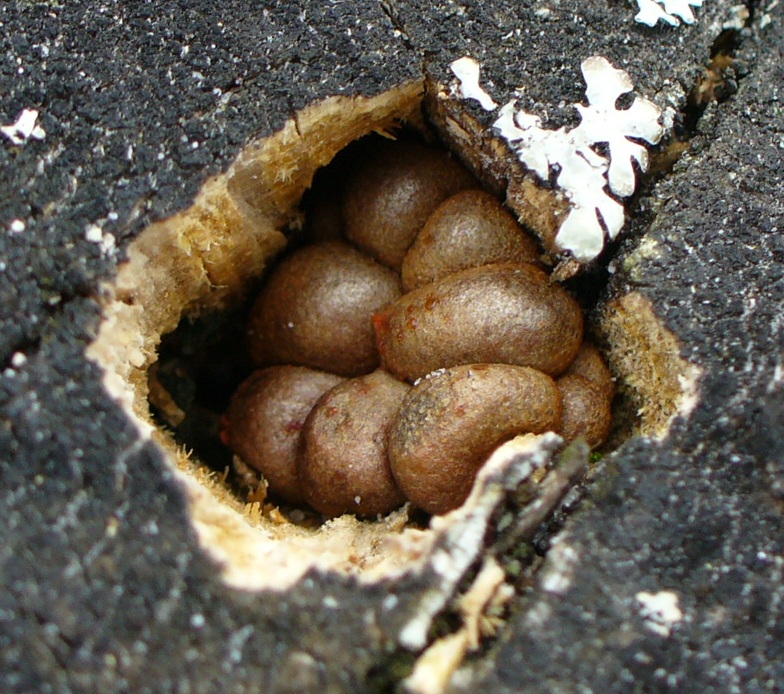
| Brown slime-mould structures, like eggs in a nest. |
 |
| Here they are emerging and also showing orange and reddish colours. |
 |
| More examples of orange structures. |
 |
| A close-up of an orange-red structure emerging from a crack in a stump. |
These four pictures are all of slime-moulds of the
Lycogala epidendrum group, a complex recently split into two species,
L. epidendrum and
L. terrestre. Microscopic examination (e.g. of the spores) is needed to separate them, so for the purposes of this post, I shall simply call them
Lycogala. In these species, the spore-bearing structures are effectively fused to form an 'aethelium' several millimetres across which can be greyish, brownish or pinkish - the 'eggs' seen above. Like
F. septica,
Lycogala can escape from dead wood via holes such as those made by beetles as well as through existing cracks and crevices. The plasmodium is reddish or pinkish as seen in the lower picture as well as near the right-hand edge of the second picture.
Now, I am no expert on slime-moulds, but I do find them intriguing - and it's quite a sight when several brightly coloured species inhabit the same piece of dead wood. However, I can recommend the excellent handbook by Ing (1999) which I am highly reliant on - it won't give you lots of colour photos (though a web search will), but it does provide simple line drawings of key structures, keys, spore measurements and so on covering the few hundred species in Britain. It's also the first comprehensive work on the British species since 1877!
Enjoy the slime!
Reference
Ing, B. (1999).
The Myxomycetes of Britain and Ireland: An Identification Handbook. Richmond, Slough.


Hi there - nice post on Slime Moulds! Used to grow then in school for Biology kids to have a look - good view of cytoplasmic streaming (and that’s not something I get to put on many people blogs!)
ReplyDeleteStrange and wonderful things to study!
Stewart M - Australia
Thanks Stewart - I was amazed to see the first three all on a single, not especially large, log.
ReplyDelete