Something that has begun to fascinate me over the last couple of years is cecidology - the study of galls. I am by no means an expert in this, though I have been drawn to joining the
British Plant Gall Society (BPGS) and last year even led a gall-hunting trip (another one's booked in for later this year). Why? Well, good question - it's a combination of factors - the diversity of forms, the fact that finding and IDing galls is often easier than finding and IDing the tiny invertebrates that create them (of course, some are made by bacteria and fungi), and the lack of knowledge about them - exactly how galls form, the ecology of many species and so on. So, when rootling around under an oak (
Quercus robur) a couple of days ago, I found a couple of old brown Knopper galls of the cynipid
Andricus quercuscalicis and decided to bring them home for a closer look...
 |
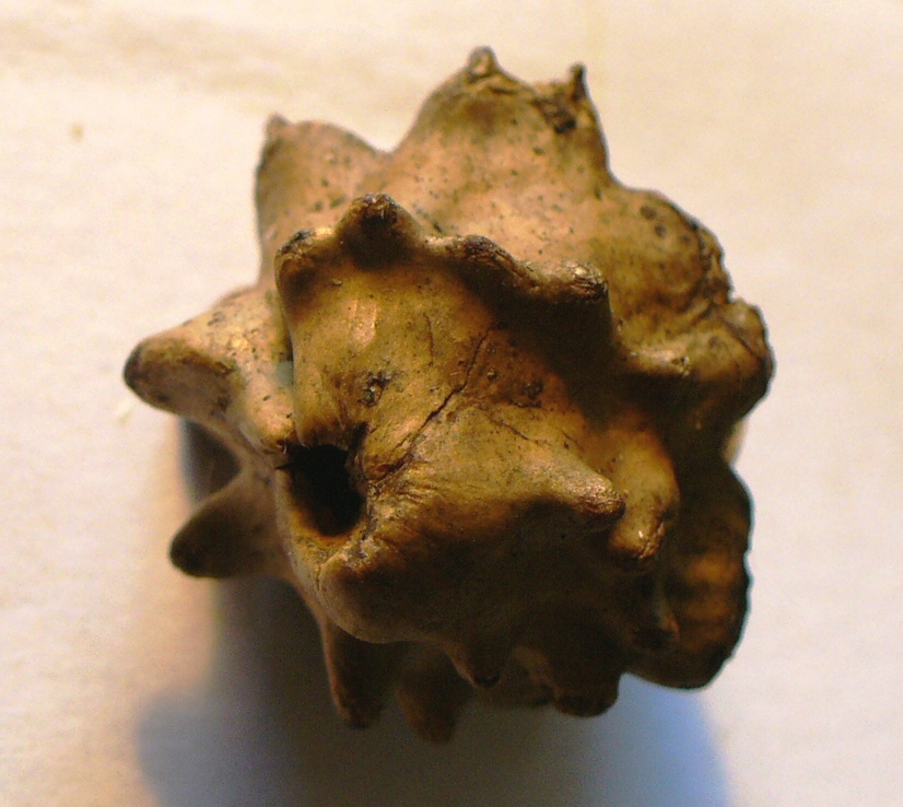
| The familiar knobbly form of a Knopper gall (green when fresh, woody when older) |
The Knopper grows as a gall on acorns forming a series of protrusions as seen above with an exit hole at the top. Cutting it open however starts to reveal a tiny world of finer structure.
 |
| One bisected Knopper... |
This shows a thick woody outer wall with an approximately conical chamber inside with thin vertical partial walls or flanges, and the exit hole at the top. Now, looking closer still...
 |
| Zooming in on the chamber and one of the protrusions. |
Above you can see some other structures - a green-grey mass near the centre and a couple of small yellowish ovals near it (more on these below). Top right is one of the projections and the section shows a pitted texture with a smooth line running through it. I have no idea what this line is - does it provide strength like a laminate or reinforcing bar? Zooming in again shows the pitted structure to be closely packed cylindrical cells.
 |
| Cells within the wall of the Knopper gall |
 |
| Egg case or larval skin? |
Even this view has a linear structure running through it - so far I've been unable to find out more, or perceive any deeper structure, but I will keep looking, and welcome any comments on this. Now, the tiny yellowish structures - I think they are either egg cases or shed larval skins (exuviae), but I could see much structure even under higher magnification so have left that for the moment, and a couple of darker structures are what I believe to be frass (insect faeces). The green-grey tufts are a little more interesting though.
 |
| Close up of tufts inside the gall |
A few possibilities sprung to mind - a lichen, or a microfungus - maybe some sort of mould growing on invertebrate detritus. A quick bit of work with a hooked pin and a microscope slide and the following appeared from the same material:
 |
| Filamentous green or blue-green alga |
 |
| Numerous fungal spores and tiny hairlike structures |
From these pictures and the general look of the tufts, I was initially pretty sure this was a lichen - a symbiotic arrangement of fungus (metabolising dead organic matter) and the 'photobiont', a photosynthesising alga or cyanobacterium associated with in. I was thinking of trying to ID the 'photobiont', but a handy comment (see below) suggests that it is a mould along with a separate alga after all (the title stays though!). I've developed quite a few questions from this one common gall, but that's one that may have been answered. Anyhow, that would have been as far as I could get for now, until I noticed a tiny flicker of movement and found this...
 |
| A tiny metallic green wasp |
 |
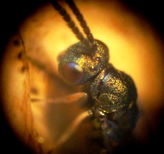
| And closer in... |
Yes, it is a tiny (2mm tops) chalcid - but not the gall-causer which is a more rotund shape and brownish in colour. This is one of its larval parasites and belongs to the family Pteromalidae. It looks very similar to some other families such as Ormyridae and Torymidae but is distinguished by the narrow pronotal collar, long stigmal vein on the wing and some features of its sculpturing. The genus is uncertain (it really needs a well prepared specimen under high magnification), but is likely to be
Cecidostiba sp. Once I've been shopping for the 'Oak Galls of Britain' book (from BPGS's publications page), I may know more.
So, that's where I've got to so far - some interesting observations (I think so, anyway) and plenty of unanswered questions, so as ever, comments and suggestions are welcome. I suspect I'll learn a lot in about a month when the New Naturalist 'Plant Galls' book comes out (already on order...) and will post updates if any of my questions are answered. Until then, enjoy the following gratuitous pteromalid close-ups - a truly lovely little parasite!
 |
| Head showing crooked antennae, red-brown eyes with ommatidia (facets) and red-brown ocelli. |
 |
| Dorsal sculpturing |
 |
| Iridescent wings and venation |
 |
| A ventral view, also showing the long stigmal vein |
 |
| A slightly broader dorsal view. |


Wow, there's a whole world in there! I've never really thought much about what's in a gall, I don't actually know much about them, but this is fascinating.
ReplyDeleteThanks - it was seeing the parasite come out that got me wondering really - two more have just emerged from the other knopper I collected, so there may be an update if I can get one under the high-power 'scope - have just ordered the relevant book mentioned in my post...
ReplyDeleteAn alga and a fungus growing intermixed doth not a lichen make! The green growth looks to me like a green mould, eg Aspergillus or Trichoderma. (The spores are wrong for Cladosporium)
ReplyDeleteWell, that is true - as mentioned in the post, this was a bit of guesswork (it would explain why I couldn't match the spores and alga well) and a mould was one of the possibilities. Happy to amend the post - as I say, comments are helpful.
ReplyDelete