Several of my post in recent months have been on the topic of 'diagnostic morphology' i.e. taking a close look at invertebrates (some common, some not) to see why they are what they are. I've looked at beetles, galls, wasps and others, but have so far written little about spiders. So, when I found a colourful little spider drowned in the garden yesterday, it seemed only fitting that its demise should be of some value - though it wrinkled once dry, the key features remain visible.
 |
| Dorsal view of the dried specimen of Enoplognatha ovata |
From the first photo, the abdomen shows the distinctive green-yellow colour with a central broad red stripe, although this is variable - the stripe is often missing with just some small dark dots instead. The carapace has a thin central dark stripe and the whole spider (excluding legs and other appendages) is about 4mm long. These features alone allow identification as
E. ovata, but there are other features such as the large divergent (spreading) chelicerae (jaws) bearing long fangs. These are seen in males of the genus
Enoplognatha, along with the 'boxing glove' palps used in reproduction. Let's look in a little more detail.
 |
| Ventral view - the palps and fangs can be seen clearly, plus the leg attachments underneath the thorax - which also shows the thin central dark line visible on the dorsal surface. |
 |
| Dorsal view of the abdomen detached from the thorax. The point of attachment is bottom left and is surrounded by small parallel stripes - probably muscle attachments. The rough ovals are the leg attachments visible through the abdomen. |
 |
| Side view of the abdomen - this shows the mosaic colour pattern which appears uniform from further away. Also, what appears to be a smooth abdomen is shown to be bristly under higher magnification. |
E. ovata is a member of the family Theridiidae, also known as the 'comb-footed spiders' and it is worth looking at some features that show its place in this group which tends to build random, tangled three-dimensional webs.
 |
| The tip of a leg showing not only the bristles, but also the 'comb-foot' that gives the family its common name. These are used to comb out silk from the spinnerets during web-building - this silk is not sticky, but entangles prey. |
 |
| The arrangement of eyes (the front is to the right). Relative to the small size of the spider, these are fairly large and protruding. Apart from the front middle eyes (which are always dark), these tend to look pearly when the spider is alive. Note the fusion of the side pairs of eyes. Also, there are a few tiny bristles present on the head. This arrangement of eyes with two short rows, the side pairs fused, is typical of the Theridiidae. |
 |
| Close-up of the fused left-hand pair showing them protruding, and the wrinkled structure where they join. Below this some faint dark lines are just visible internally which may be nerve fibres running from the eyes. |
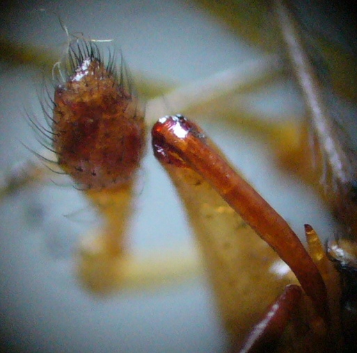
Lastly I'd like to look at the appendages attached to the underside of the head - the chelicerae and palps.
 |
| In the left, the male's oval palp. On the right the large fang of one of the chelicerae. In other genera within the Theridiidae, the chelicerae are no more than medium-sized. Here the central channel is just visible running through the centre of the fang - this family of spiders has a fast-acting venom which, though they can't bite humans, quickly disables its invertebrate prey. |
 |
| The male palp.The structure varies by species and receives spermatozoa from the testes. It is then used to transfer sperm to the female. In adult male spiders, the palps are the most important feature needed for identification to species (where this is not otherwise straightforward). In females, it is the epigyne, the area around the genital opening which receives sperm from the palps. Here note the bulbous end segment of the palp forming a 'cymbium' within which the palpal organs (the reddish protrusions etc) are located. |
As in previous posts of this type, I hope this helps clarify the importance and function of fine structure - sometimes it is required for identification, but in any case is interesting as it illustrates how 'form and function' are inextricably linked.
References
Jones, D. (1989).
A Guide to Spiders of Britain and Northern Europe. Hamlyn, London.
Roberts, M.J. (1995).
Spiders of Britain and Northern Europe. HarperCollins, London.


Cool morphology shots.
ReplyDeleteThanks - glad you liked it - more morphologu coming up soon; moths, ground beetles and hoverflies all vaguely planned...
ReplyDelete