Having looked at the
abdomen and
cephalothorax, it's time for the third and final instalment of tarantula anatomy based on the features visible on a moulted skin.
 |
| Ventral view of the sternum showing articulations with legs and chelicerae |
Although legs and chelicerae are the most prominent appendages and found on the cephalothorax, I want to start with the spinnerets found at the end of the abdomen. Even though this is not a web-weaving spider, it still needs to produce silk e.g. to form eggs cocoons (this is a female), and does so from the paired spinnerets. Each spinneret is linked to silk glands and is a segmented structure to allow the silk to be manipulated as it is extruded.
 |
| One of the spinnerets - note the segmentation and longitudinal groove in the last segment. |
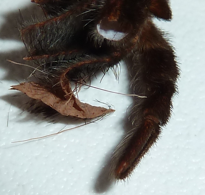
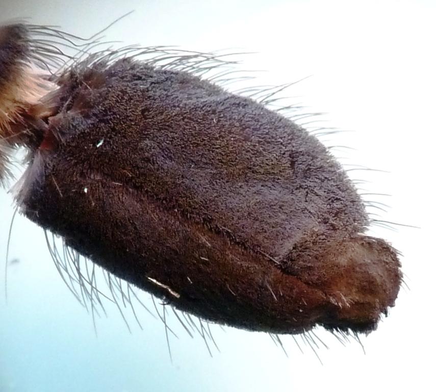
Moving forward, a key feature is of course the presence of eight legs - the source of much arachnophobia. These are clearly jointed and are dark with orange-red bands giving this species its common name. However, the part I want to look at is the end of the final segment or tarsus - the spider's 'foot' if you like. Whereas the rest of the leg (and much of the spider) is covered in a variety of bristles, the foot-pad is covered in tiny, short hairs giving it a soft, velvety appearance. More importantly, this also means that the pad has a large surface area, aiding grip - much like the almost fractal convoluted ridges on a gecko's foot (maybe more on that another time - there are electrostatic effects involved which are fascinating), as well as a pair of small claws.
 |
| The underside of the tarsus showing a velvety covering of small hairs. |
 |
| Tip of the tarsus with the tiny claw (one of a pair) indicated. |
Moving further forward, the next appendages are of course the mouthparts, in particular the fang-bearing structures called chelicerae.
 |
| Ventral view of the chelicerae showing fangs. |
The chelicerae are articulated and can flex the fangs forward to grip prey as the fangs are hardened and sharply pointed. As the spider is not strongly venomous, it chews rather than sucks its prey, using the fangs to grip and press food against a line of smaller teeth on the front edge of each chelicera.
 |
| Chelicera showing fang, smaller teeth and flat inner surface. |
The inner edges of the chelicerae are flat and relatively hairless as they fit closely against each other, with fringing hairs around the edge as shown above (these may be used to stridulate i.e. produce sound when rubbed together, at least in some species). Looking inside a chelicera, there is evidence of the mobility of this structure required in order to chew and manipulate food - connective tissues can be seen which would have been attached to muscles that move the chelicera and fang.
 |
| View into a chelicera showing the bases of small teeth, plus the remnants of connective tissue used to move the appendage and fang. |
That brings me to the end of my trilogy of tarantula anatomy posts. There are of course many other structures in a live spider, but the skin is easy to manipulate and can be dissected without harming the spider which has left it behind. So, if you are a tarantula-keeper, why not have a look the next time your spider moults - there are many interesting structures the closer you look.


No comments:
Post a Comment