The title of a lost H.P. Lovecraft tale? Nope, though it isn't every Christmas party that you get handed a box containing a mummified corpse...
 |
| A mummified common lizard, Zootoca vivipara |
Actually, this isn't as odd as it sounds - the lizard was found by friends while taking up an old carpet, and might be a casualty of one of their cats that subsequently dried out. As you'll see later, it isn't perfectly preserved... Anyhow, the first thing I did was measure it - at approximately 40mm in length including tail, it was newly born as the common (or viviparous) lizard (
Zootoca vivipara, previously
Lacerta) is 37-44mm long at birth with the tail no more than about half the overall length (Beebee & Griffiths 2000, Inns 2009). Then, as ever, I took a closer look...
 |
| Dorsal view of the Z. vivipara specimen |
As you can see, the skin/scales of the head is largely intact and the scale pattern remains clearly visible, a feature that can be useful for identification, especially in areas (unlike the UK) with a wide range of superficially similar species. As is typical for young common lizards, the colour is dark, though this could be an artefact of death and drying, and zooming in on the abdominal scales gives a hint of the colour that develops after a few weeks. The dense, overlapping arrangement of dorsal scales (with 'keels') is typical of lacertids.
 |
| Dorsal abdominal scales of young Z. vivipara - dark but with a hint of the metallic greenish-brown-bronze colour (including some gold flecks) that typically develops after a few weeks (Beebee & Griffiths 2000) |
Turning the specimen over, further structures can be seen. As well as skeletal features such as the ribs, some of the ventral scales (or 'plates') are intact (though not the collar area beneath the neck, nor the femoral pores beneath the thigh), and show the development of an iridescent greenish colour. The scales are not simple plates of keratin like the 'scutes' of crocodilians, but are outfoldings of the skin itself that are each covered in a tough waterproof layer and contain both an osteoderm (internal bony plate) for structural integrity and a layer of chromatophores - cells that produce colour - just below the outer epidermis.
 |
| Ventral view of young Z. vivipara |
 |
| Ventral scales/plates of young Z. vivipara |
Turning the specimen on its side then allows the skull to be investigated as the skin is missing. You can immediately see the teeth (more below on this) - as is usual for lizards, they are conical and all approximately the same size - and show that the young common lizard is well equipped to begin feeding (on anything small enough to catch, including aphids!) as soon as it is born. It's also worth taking note of the overall structure as lizard skulls are rarely represented in books on animal signs (unlike mammal skulls, a range of which tend to be illustrated) or other resources such as guides to the contents of owl pellets (presumably owls rarely if ever eat them).
 |
| Side view of the head of young Z. vivipara. |
However, I did notice something towards the rear of the skull - you can just see it in the photo above as a pair of little white dots in the left-most of the three main cavities. Time to switch to a higher magnification...
 |
| Small Fungi growing on the head of Z. vivipara |
...and another kingdom of organisms as these are clearly Fungi. These structures are around 0.5mm long, with the white 'blob' around 0.1mm in diameter. I can't begin to describe how fiddly it was removing these to make a slide, but I did (tiny tweezers) and decided to see if I could identify the fungus.
 |
| Fungus from Z. vivipara (the arrows indicate the length of one of the stalk cells) |
This is clearly a spore-mass on a stalk (one cell thick, cell walls indicated by arrows). The white colour of the spore-mass in the first photo is an artefect of lighting and my camera; it is actually a pale yellowish-brown - darker in the second photo where it has been compressed. Consulting Ellis & Ellis (1998) it soon became clear this wasn't a specialised 'bone-eating' fungus but was more likely to be one of the moulds (Ascomycetes) that can be found on a wide range of organic matter. Given the overall form/size and the more-or-less round and transparent spores (some of which appear slightly warty), this is probably
Eurotium and/or
Aspergillus, although the faint equatorial groove of
Eurotium isn't visible in the photos. 'And/or'? you may ask... well...
 |
| Cleistothecium (spore mass) of Eurotium /Aspergillus |
 |
| Warty spores of Eurotium/Aspergillus indicated by arrows. Others are not in focus. |
The taxonomy of some fungal groups is complex because different (asexual 'anamorph' and sexual 'teleomorph') stages have been given different names, and in some cases there are several anamorphs, all with separate names. This is case here with
Eurotium being the teleomorph and
Aspergillus the anamorph - both of which may be found together, and often are. I could expand on this, but fortunately don't need to - as of January 1st 2013, any one fungus will have a single name covering all stages/'morphs', and details of how this will work are given in
Hawksworth (2011). As far as this specimen is concerned, taxonomic considerations aside, it does mean that it is mouldy and therefore not perfectly mummified. I have it in a dry container so it may surrvive, or I might find it turns into a clean skeleton. Certainly this eruption from the scales shows it's still active.
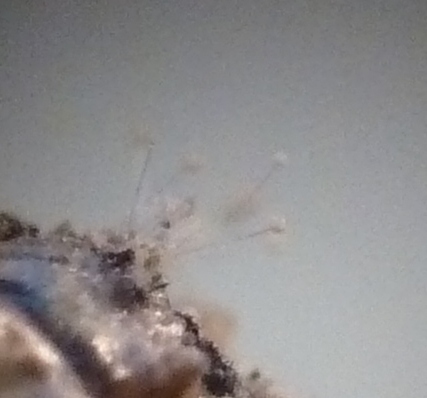
 |
| 'Tuft' of Eurotium/Aspergillus growing from the skin of Z. vivipara |
Now, back to the lizard... following a reader's comment, I returned to the specimen and had a better look at the teeth. I also Googled and was surprised to find few images showing lizard teeth in detail - presumably they exist, but not on my bookshelves. As mentioned above, the structure is fairly uniform (unlike us mammals with canines, incisors, molars with varying numbers of cusps and so on), but that is a slight oversimplification.
 |
| Cheek teeth of juvenile Z. vivipara |
 |
| Front teeth of juvenile Z. vivipara |
Ignoring the fungal structures this time, the cheek teeth are clearly more-or-less cylindrical with a conical point; they are also transparent showing what I presume is the pulp cavity as a whitish central mass. However, the front teeth are curved backwards and more 'fang-like' as might be expected in order to catch and hold prey. Although showing little obvious differentiation, a search of the literature online indicates (and I'm way out of my comfort zone here!) that lacertid teeth can be categorised as premaxillary (apparently of taxonomic and palaeontological importance with 7 in
Zootoca), maxillary and dentary according to their position and associated jawbones (
Arribas 1998). This is where I'll stop on dentition, but if any reader would like to expand on this in the comments (e.g. whether the different tooth groups differ structurally), please feel free!
Lastly, I can't finish this post without mentioning reptile conservation in the UK. The common lizard is widespread but has declined, especially in the south, due largely to development pressure and the loss of brownfield sites. Because of this it is now a
UK BAP (Biodiversity Action Plan) species. It seems clear that habitat loss needs to be tackled (although with a government that seems to want to relax, rather than tighten, planning laws it is unclear how this will happen), but there is good advice in Gent & Gibson (2003) and Edgar
et al. (2010). Rather than repeating what they have said, or putting my campaigning hat on, I'd just like to highlight the importance of collecting (and sending in to Biological Records Centres) reptile species records, especially as part of systematic surveys though
ad hoc records are valuable too, and undertaking practical conservation work to improve habitat quality.
References
Arribas, O.J. (1998). Osteology of the Pyrenean Mountain Lizards and comparison with other species
of the collective genus
Archaeolacerta Mertens, 1921 s. l. from Europe and Asia Minor (Squamata: Sauna: Lacertidae).
Herpetozoa 11(1/2): 47-70.
Beebee, T. & Griffiths, R. (2000).
Amphibians & Reptiles. HarperCollins, London.
Edgar, P., Foster, J. & Baker, J. (eds.) (2010).
Reptile Habitat Management Handbook. ARC, Bournemouth.
Ellis, M.B. & Ellis, J.P. (1998).
Microfungi on Miscellaneous Substrates (2nd ed.). Richmond, Slough.
Gent, T. & Gibson, S. (eds.) (2003).
Herpetofauna Workers' Manual (revised reprint). JNCC, Peterborough.
Hawksworth, D. (2011). A new dawn for the naming of fungi: impacts of decisions made in
Melbourne in July 2011 on the future publication and regulation of
fungal names.
Mycokeys 1: 7-20.
Inns, H. (2009).
Britain's Reptiles and Amphibians. WildGuides, Old Basing.


Very interesting Dave. I thought you would like the lizard skeleton for your collection of 'interesting things', I never thought you would go to such lengths and I would learn so much about Z.vivipara.
ReplyDeleteThe photo's are excellent.
Laura
Brilliant stuff - I would have used the microscope to look in detail at the teeth, but I suppose fungi are interesting to some (smiley). Great photos!
ReplyDeleteGood point Darren, 'tis done!
ReplyDelete